Modern magnetic resonance imaging techniques allow not only a high-resolution imaging of brain structures but also an image of the connectivity, i.e. the fiber connections between the individual areas in the brain. This is particularly important when planning surgeries that take place in the vicinity of critical fiber connections such as the visual pathway or motor pathways. In neurosurgery at Inselspital, fiber tracking is used for special operations to ensure maximum patient safety and the best possible outcome after the operation.
How was fiber tracking technology developed?
Fiber tracking or diffusion tensor imaging refers to the visualization of fiber bundles that connect functional centers in the brain (e.g. motor speech production and speech comprehension). This visualization can either be included in preoperative access planning or displayed directly in the surgeon's operating microscope during the operation.
The beginnings in the 1980s
The first publications on diffusion tensor imaging were made as early as the 1980s *, * and the technology has been continuously improved since then.
The method makes use of an effect that flowing water exerts on magnetic resonance imaging. In a water glass, for example, water can flow uniformly in all directions. There is no preference for any direction. Thus, during a magnetic resonance imaging scan of the glass of water, one could measure what's called isotropy at every point, meaning direction independence. In the brain, water is found in blood vessels and also in tissue within the nerve cells and the connective tissue cells surrounding them. In larger fiber bundles, water flows preferentially in the direction of the fibers. This is referred to as focal anisotropy, i.e. directionality. This can be determined by special magnetic resonance imaging measurements for each point in the brain and is described by an "eigenvector".
Further development of the method
Further development of the methodology was the color-coding first shown by Sinisa Pajevic and Carlo Pierpaoli in 1999, in which fibers in the head-foot direction are shown in blue, fibers in the left-right direction are shown in red, and fibers in the front-back direction are shown in green *. This made it possible, for example, to show the pyramidal tract as a strong, deep blue structure that connects the nerve cells in the brain mantle with the spinal cord, which is essential for the movement of arms and legs.
Modern high definition fiber tracking (HDFT)
Despite these advances, limitations in imaging resolution persisted. In 2012, high definition fiber tracking (HDFT) was developed, based on the previous technology *. HDFT made it possible to correctly display crossing fiber connections with a few exceptions and to determine the origin and target point of the fiber connection more precisely. For today's neurosurgical application, this new method has opened up fundamental possibilities in surgical planning, as HDFT could now also accurately depict the exact course of the cranial nerves, which was previously only possible to a limited extent *. Similarly, this new methodology can now also be used to precisely depict important fiber bundles such as the visual pathway. This makes interventions near these functional centers much safer for the patient.
How is fiber tracking used at Inselspital?
When brain tumors are located near functional pathways, we use fiber tracking as a standard part of our surgical planning. This mainly concerns tumors close to the speech, visual, or motor centers. Prior to surgery, the visualization of the pathways is individually created for each patient, and access to the tumor is planned accordingly.
The goal is radical removal, even for tumors in important functional areas of the brain. Together with intraoperative neurophysiology monitoring (IONM), we achieve maximum radicality for our patients with minimal invasiveness and important functional preservation.
The planning is based on data calculated by a computer, which in principle can also be influenced by technical errors. In addition, the resolution of the underlying magnetic resonance imaging (MRI) is limited so that only bundles of many thousands of nerve fibers can be examined together rather than individual nerves. Despite modern imaging techniques such as HDFT, unexpected breaks in the reconstructed fibers continue to occur at intersections and near fibers that change course in a confined space. Picking out the relevant fibers, checking their plausibility, and preparing them for surgical planning is the task for a IONM specialist.
In order to make operations even safer for patients in the future, research and further development of innovative techniques such as fiber tracking form an independent research focus at Inselspital.
Fiber tracking in clinical application
- Planning of the fiber tracks
If an operation is to be performed with the aid of fiber tracking, the specially required DTI sequences and T2-weighted image sequences are created and merged in advance in a 3 Tesla magnetic resonance imaging (MRI). Using an automatic algorithm, artifacts (eddy current artifacts and motion artifacts) are automatically eliminated. To calculate the desired fiber bundles, a region of interest (ROI) is now superimposed over the relevant anatomical landmarks in the T2-weighted MRI imaging. Furthermore, one selects the parameters for fractional anisotropy and fiber bundle length.
For example, to calculate the pyramidal tract, the regions of interest are plotted along with the imaging anatomical landmarks of the pyramidal tract on the T2-weighted images: gyrus precentralis, capsula interna, and parts of the pedunculus cerebri. With the correctly chosen adjustment parameters, a homogeneous fiber bundle is obtained, which represents the course of the pyramidal tract.
")
Regions of interest (ROI). In T2-weighted MRI images, the ROIs are marked to calculate the pyramidal tract. The pyramidal tract (corticospinal tract) originates in the precentral gyrus (left image) and runs via the cerebral crus (right image) into the spinal cord. Bild: Nowacki et. al. 2018, JNS - Fiber tracking in tumor surgery
If a tumor is located near a vital fiber bundle, fiber tracking is necessary to ensure maximum patient safety. Often, the boundaries between healthy brain tissue and tumor tissue cannot be clearly delineated. The exact course of the fiber bundles in relation to the tumor provides the surgeon with important additional information that is not visible to the naked eye.
Together with other techniques such as intraoperative neuromonitoring, the surgeon has several options to ensure that no critical structures are injured during surgery. In particularbfiber connections which are necessary for basic human functions such as speech, vision, and movement are protected with great effort from unintended damage during tumor surgery.

Tumor surgery. (Left) The tumor to be removed (T) is in close proximity to the arcuate fasciculus (AF). The middle image shows the so-called voxels of interest (VOI), which were used to calculate the fiber bundle. On the right is the course of the arcuate fasciculus with Broca's center (B) and Wernicke's area (W) at each end of the fiber bundle. Bild: Stieglitz et. al. 2012, Neurosurgery 
Eloquent areas. This image shows a tumor (orange) in the middle of functionally important areas: corticospinal tract (blue), optic tract (yellow) and arcuate fasciculus (green) Bild: Universitätsklinik für Neurochirurgie, Inselspital Bern © CC BY-NC 4.0 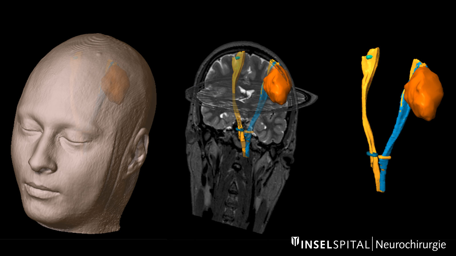
Corticospinal tract. The corticospinal tract (yellow and blue) is shown here as a fiber tract of motor function in relation to a tumor (orange). Bild: Universitätsklinik für Neurochirurgie, Inselspital Bern © CC BY-NC 4.0 
Visual pathway. This illustration shows how the visual pathway (optic tract, green) runs around the ventricular system (purple). Bild: Universitätsklinik für Neurochirurgie, Inselspital Bern © CC BY-NC 4.0 - Fiber tracking for deep brain stimulation
Essential tremor
In essential tremor, we use fiber tracking to visualize the dentatorubrothalamic tract (DRTT) to plan stimulation of functional pathways. This complements traditional purely anatomical planning. The DRTT is located in the posterior subthalamic area (PSA) and is one of the target points in deep brain stimulation (DBS) for the treatment of essential tremor *. Accurate localization is essential in surgical planning to allow precise stimulation of the tract. Fiber tracking can be used to visualize the tract and optimally determine the target points for DBS electrode implantation.

Deep brain stimulation for tremor disease. The left image shows the visualization of the dentatorubrothalamic tract for planning the correct electrode implantation (red) for deep brain stimulation. The right image shows the exact target localization so that the stimulation can take place directly next to the DRTT. Bild: Universitätsklinik für Neurochirurgie, Inselspital Bern © CC BY-NC 4.0 Depression

Deep brain stimulation for depression. The left image shows a 3D reconstruction of the right (red) and left (green) medial forebrain bundle. On the right image, the target points for electrode placement prior to deep brain stimulation are marked as white dots. Bild: Fenoy et. al. 2016, J Affect Disord Another clinical application for fiber tracking at Inselspital is deep brain stimulation (DBS) therapy for severe cases of depression that do not respond to standard therapy. In medical studies, it has been shown that stimulation of the superolateral part of the medial forebrain bundle has a positive therapeutic effect in refractory depression *. In order to correctly identify the fiber bundle and determine the target coordinates for DBS, fiber tracking is necessary prior to the procedure.
")

-
Taylor DG , Bushell MC. The spatial mapping of translational diffusion coefficients by the NMR imaging technique. Phys Med Biol. 1. April 1985;30(4):345–9.
-
Merboldt K-D, Hanicke W, Frahm J. Self-diffusion NMR imaging using stimulated echoes. Journal of Magnetic Resonance. Magn Reson Med. 1991 Jun;19(2):233-9.
-
Pajevic S, Pierpaoli C. Color schemes to represent the orientation of anisotropic tissues from diffusion tensor data: application to white matter fiber tract mapping in the human brain. Magn Reson Med. September 1999;42(3):526–40.
-
Fernandez-Miranda J, Pathak S, Engh J, Jarbo K, Verstynen T, Yeh F, YWang Y, Mintz A, Boada F, Schneider W, Friedlander R. High-Definition Fiber Tractography of the Human Brain: Neuroanatomical Validation and Neurosurgical Applications. Neurosurgery. Aug 2012;71(2):430-453
-
Yoshino M, Abhinav K, Yeh F, Panesar S, Fernandes D, Pathak S, Gardner P, Fernandez-Miranda J. Visualization of Cranial Nerves Using High-Definition Fiber Tractography. Neurosurgery. 2016;146-165.
-
Nowacki A, Debove I, Rossi F, Schlaeppi J, Petermann K, Wiest R, Schüpbach M, Pollo C. Targeting the posterior subthalamic area for essential tremor: proposal for MRI-based anatomical landmarks. Journal of Neurosurgery. 2018;1-8.
-
Fenoy AJ, Schulz P, Selvaraj S, Burrows C, Spiker D, Cao B, et. al. Deep brain stimulation of the medial forebrain bundle: Distinctive responses in resistant depression. J Affect Disord. Oktober 2016;203:143–51.