Neuronavigation is a computer-assisted procedure that is used to navigate precisely and safely in the brain or spine. It combines imaging procedures such as CT or MRI scans with special software that makes it possible to create three-dimensional models of the anatomical conditions and use them during the operation. In addition to classic neuronavigation for the head, neuronavigation has also become established for spinal surgery in recent years. Whereas it used to be necessary to take repeated X-rays during the operation, the anatomical conditions can now be displayed precisely on the screen and instrument guidance can be visualized. This increased accuracy allows even complex procedures to be performed on the entire spine.
What are the advantages of spinal neuronavigation?
Increased accuracy
A working group led by Shin and colleagues conducted a systematic analysis of spinal navigation for screw implantation in the spine *. In this study, navigated and non-navigated screw implantations were compared. The correct position of implanted screws and the complications of both procedures were examined. A comparison of 20 studies showed that the risk of screw misplacement in navigated operations was only 6%, while the risk in conventional operations was 15%. Furthermore, there were no neurological complications in the navigated screw operations.
Other studies, such as those conducted by the working group of Richter et al * or Fichtner et al *, also showed a safe transpedicular screw position with greater accuracy using spinal navigation.
Lower radiation exposure
The navigation system can also reduce the radiation dose to a minimum, both for the patient and for the surgical team *.
How is spinal neuronavigation used?
There are different spinal neuronavigation systems:
For example, a 3D image of the spine obtained before the operation using computed tomography (CT) can be fused with an intraoperative 3D X-ray image of the spine. Software algorithms fuse the three-dimensional matrix of the X-ray image with the images of the preoperative CT data set. This was frequently used in the past.
Now, thanks to further development of the navigation software, it is also possible to compare the surface of the patient's bone directly with preoperative CT images. In this case, the bone of the vertebral body is compared with the CT image and the data is transferred intraoperatively to the surgical monitor. This makes it possible for the surgeon to track the position of his instruments and the position of inserted screws in real time on a three-dimensional data set of the spine.
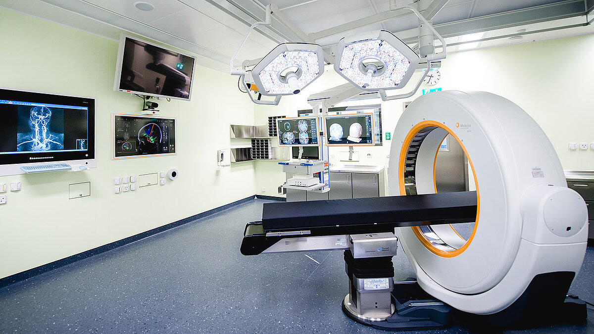
At Inselspital: Intraoperative computed tomography (iCT)
CT-based spinal navigation with intraoperative CT (Airo®) is used as standard in our Department of Neurosurgery. After intubation of the patient and positioning on the operating table, a reference star is first attached to the patient's spine before an intraoperative CT is performed for navigation. Communication between the CT and navigation software means that the images obtained in this way can be made available directly for the operation. They form the basis for high-precision navigation accuracy. After implantation of the screws or implants, a second intraoperative CT scan is performed during anesthesia for quality assurance. Any screw misalignment can therefore be corrected immediately. Our patients can thus be spared subsequent revision operations.
This procedure is also standard at our clinic for minimally invasive spinal surgery.
-
Shin BJ, James AR, Njoku IU, Härtl R. Pedicle screw navigation: a systematic review and meta-analysis of perforation risk for computer-navigated versus freehand insertion. J Neurosurg Spine. 2012;17:113-122.
-
Richter M, Cakir B, Schmidt R. Cervical pedicle screws: conventional versus computer-assisted placement of cannulated screws. Spine (Phila Pa 1976). 2005;30:2280-2287.
-
Fichtner J, Hofmann N, Rienmüller A et al. Revision Rate of Misplaced Pedicle Screws of the Thoracolumbar Spine-Comparison of Three-Dimensional Fluoroscopy Navigation with Freehand Placement: A Systematic Analysis and Review of the Literature. World Neurosurg. 2018;109:e24-e32.
-
Schuetze K, Eickhoff A, Dehner C, Schultheiss M, Gebhard F, Richter PH. Radiation exposure for the surgical team in a hybrid-operating room. J Robot Surg. 2019;13:91-98.