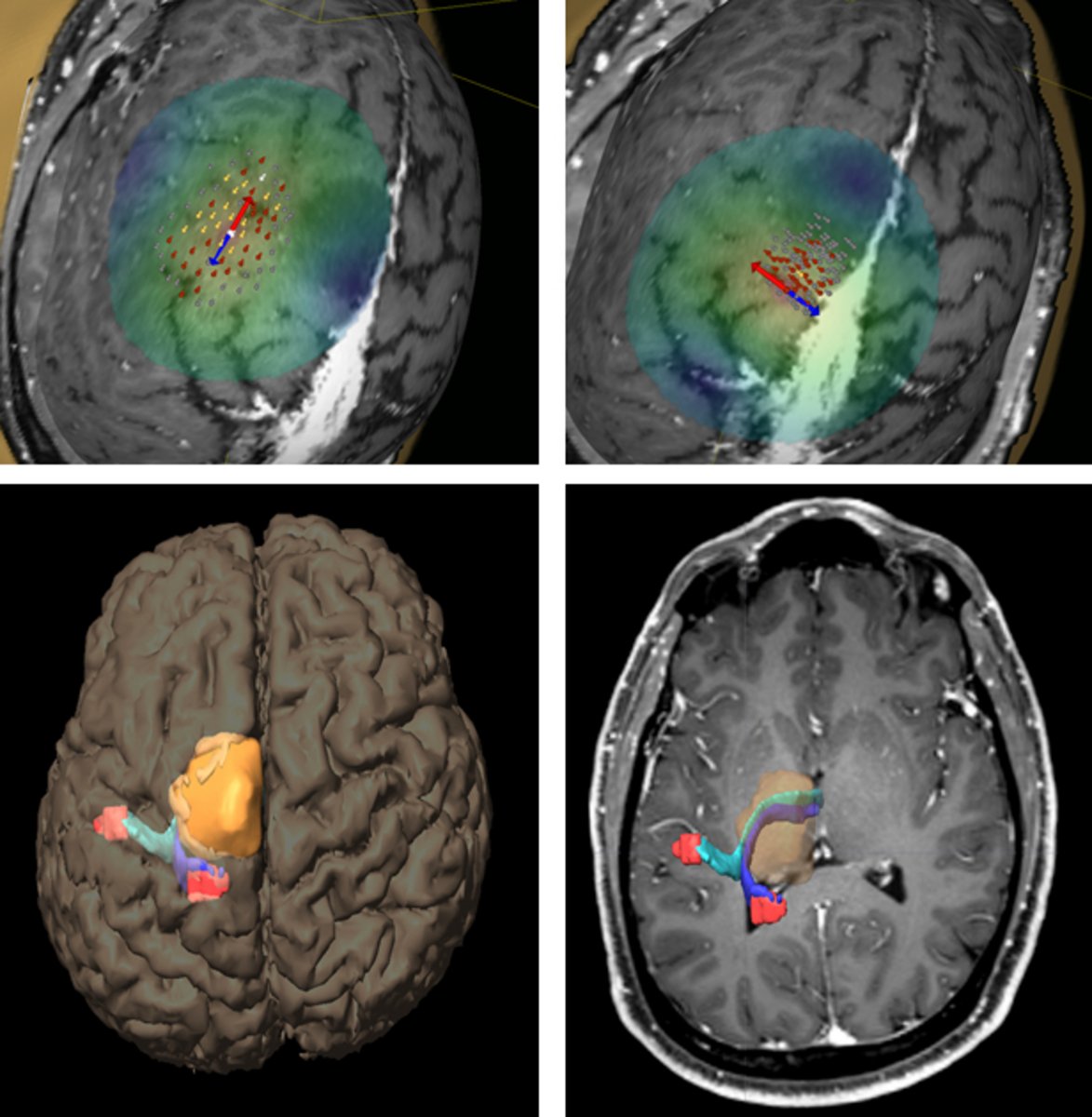
If the neurosurgeon knows precisely where functional areas of the brain are located in advance of brain surgery, he can plan the procedure safely and minimize the risk of possible consequential damage. Such brain mapping is possible with the help of navigated transcranial magnetic stimulation (nTMS). This is a non-invasive, neurophysiological technique that allows important functions in the brain to be localized with millimetre precision, as if on a map. For example, it shows exactly where the area of the brain responsible for motor function is located and how far away from a tumor it is.
How does transcranial magnetic stimulation work?
TMS is a non-invasive procedure in which nerve cells in the brain are stimulated from the outside. In TMS, a magnetic coil is placed on the patient's scalp. The coil generates rapidly alternating magnetic fields. This triggers a short electrical impulse in the brain. This impulse activates the connection between the part of the brain that controls movement, the motor cortex, and the muscles. A stimulus is transmitted via the cells of the muscle in the form of an action potential, which causes the muscle to contract.
By registering the patient's head in a 3-dimensional space and integrating the data into the neuronavigation system, the location of the coil on the surface of the head can be depicted on the patient's MRI. This makes it possible to show precisely where the brain is being stimulated. For example, if the hand area of the motor cortex is stimulated, the fingers move.
The motor-evoked potentials (MEP) generated at the time of stimulation can be measured, visualized and analysed. By repeatedly stimulating different areas of the scalp and recording the muscle responses, neurologists or neurosurgeons can precisely visualize which areas of the brain are responsible for certain movements or functions.
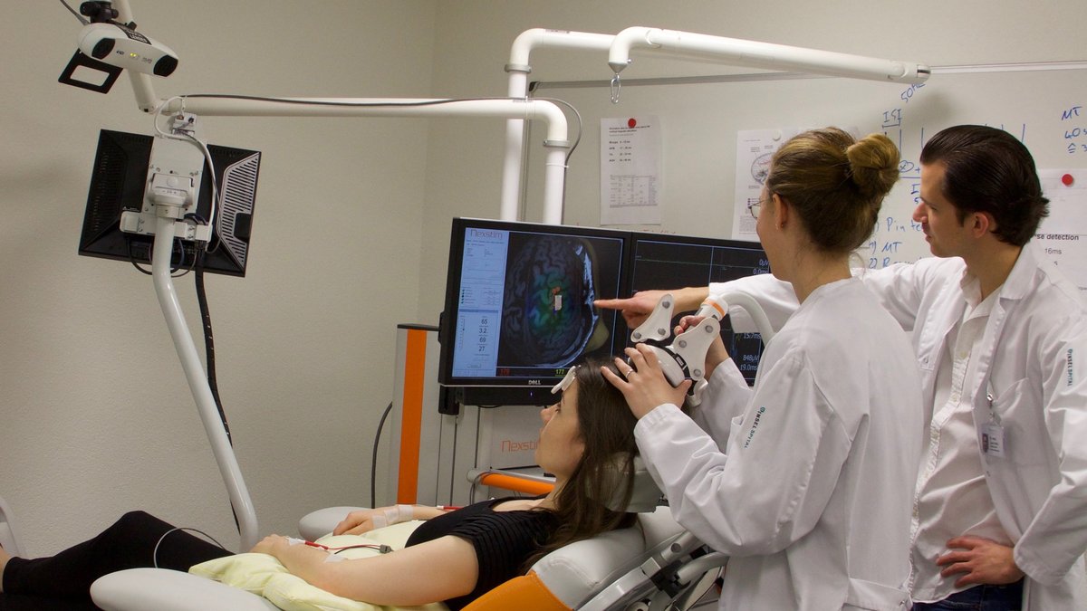
How does navigated transcranial magnetic stimulation work?
At the beginning of the TMS procedure, you will take a seat in a special chair. Your current image from a magnetic resonance imaging (MRI) scan will be loaded into the TMS device. Three points are determined as coordinates. These points are located on your head using an infrared camera and reference points. This allows for precise stimulation down to the millimeter.
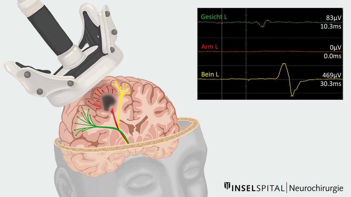
Depending on which motor center is affected in your case, adhesive electrodes will be placed on the corresponding muscles. Through electromyography (EMG), the electrical muscle activity can be measured and graphically represented. Your main task during the examination is to relax. The magnetic coil is then used to locate the motor center of the muscles under investigation and to create a corresponding map, which assists the neurosurgeon in planning the tumor removal *.
Better predictions after surgery
In rare cases, after an operation that has taken place in close proximity to the motor center, there may be a restriction in strength. In such cases, we can predict how quickly recovery will occur using the same examination method. If good responses can be derived from the arms and legs, the patient will recover quickly *.
We at Inselspital are actively involved in the further development of these innovative methods as part of international research projects and studies.
-
Krieg SM, Lioumis P, Makela JP, et al. Protocol for motor and language mapping by navigated TMS in patients and healthy volunteers; workshop report. Acta neurochirurgica. 2017;159:1187-1195.
-
Seidel K, Häni L, Lutz K, Zbinden C, Redmann A, Consuegra A, Raabe A, Schucht P. Postoperative navigated transcranial magnetic stimulation to predict motor recovery after surgery of tumors in motor eloquent areas. Clin Neurophysiol. 2019 Jun;130(6):952-959.