Neuroendoscopy plays a crucial role in the surgical treatment of certain neurological conditions and is therefore regularly utilized. Endoscopes are specialized instruments connected to a camera system and a light source via an optical lens. They provide excellent, high-resolution images from deep-seated surgical areas or cavities such as brain ventricles or cysts through tiny access points. Simultaneously, micro-instruments can be introduced through endoscopy systems to perform minimally invasive surgical procedures under complete visual control.
When was endoscopy developed?
The first endoscopic procedures on the brain took place as early as the beginning of the 20th century *. However, standardized use in clinical practice only became prevalent at the beginning of the 21st century. Reasons for this included the inadequate miniaturization of endoscopy and the insufficiently powerful light sources. By the end of the 1980s, significant technological improvements and the introduction of endoscopic working instruments (such as lasers, bipolar electrodes, and mini grasping forceps) led to a notable advancement of this technique *. Nowadays, endoscopy is indispensable for certain procedures in the neurosurgical clinical routine.
How are endoscopes used in neurosurgery?
In neurosurgery, endoscopes are used on the one hand to support microsurgical operations, for example to gain a better insight into areas that are difficult to see. On the other hand, special procedures, particularly in the area of the brain ventricles, can be performed purely neuroendoscopically. The surgeon has various tools at his disposal, such as an endoscopically usable laser, bipolar electrodes or grasping forceps.
If necessary, endoscopes can be used in combination with a neuronavigation system in order to gain additional orientation security in addition to visual control.
Which operations are suitable for endoscopic procedures?
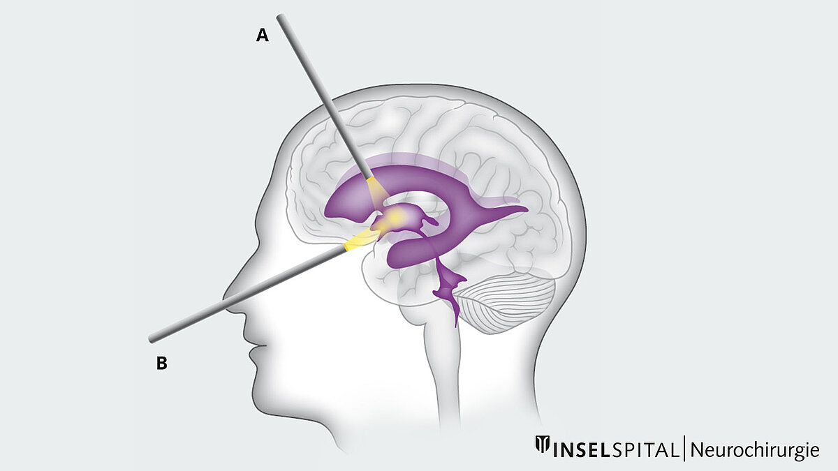
Neuroendoscopic surgical procedures are routinely used in the ventricular system and the skull base. Tumors in the ventricular system often lead to a blockage of cerebrospinal fluid (CSF) drainage, resulting in the enlargement of the brain ventricles. This enlargement creates a more favorable working environment for the surgeon *. Particularly, the lateral ventricles and the third ventricle are well accessible for neuroendoscopic surgical intervention.
Neuroendoscopy at Inselspital
Generally, we plan surgeries and the use of various surgical techniques individually for each patient. The appropriate surgical procedure will be precisely explained during the consultation in our clinic and during the pre-operative informed consent discussion.
For the listed medical conditions below, either a purely neuroendoscopic or a neuroendoscopically-assisted operation may be appropriate.
Hydrocephalus occlusus
In most cases, treatment of hydrocephalus consists of artificial CSF drainage via a ventriculoperitoneal shunt.
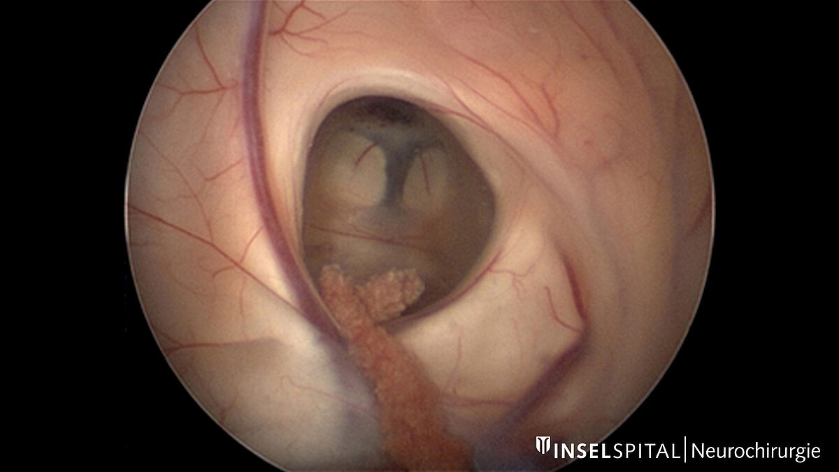
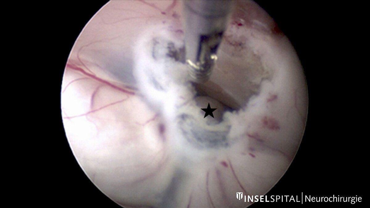
However, individual forms of hydrocephalus allow the CSF circulation to be restored alternatively via a full-endoscopic procedure, thus avoiding the implantation of a drainage system. This can be considered, for example, in cases of hydrocephalus occlusus, which can be caused by aqueductal stenosis, cysts, or tumors. A refined solution in such cases is the endoscopic third ventriculocisternostomy (ETV), in which the floor of the 3rd ventricle is opened endoscopically, thus allowing CSF drainage to the external CSF spaces.
A much rarer form of hydrocephalus may be isolated congested temporal horns of the lateral ventricle, which may occur, for example, secondarily as a complication after tumor surgery. Unfortunately, there is no standard therapy for this rare condition, but an endoscopic approach is a treatment option in some cases *.
Arachnoid cysts
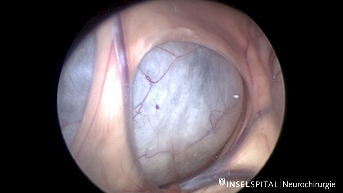
Arachnoid cysts are benign cystic changes of the middle meninges arachnoid, which are primarily asymptomatic and rarely require surgical treatment. Symptomatic cysts or cysts that become larger may require what is known as cyst fenestration. Depending on the localization and radiological findings, this can be performed purely endoscopically and minimally invasively. Suprasellar arachnoid cysts are particularly suitable for this procedure if they lead to a hydrocephalus. The dilated ventricles allow good access and the possibility of large fenestration of the cyst, which significantly reduces the risk of recurrence. Study data showed that repeated fenestration of suprasellar cysts is necessary only in the rarest of cases *.
However, the neuroendoscopic surgical procedure is not limited to suprasellar arachnoid cysts but can also be used for cysts in the middle and posterior fossa if sufficient access is available *.
Colloid cysts
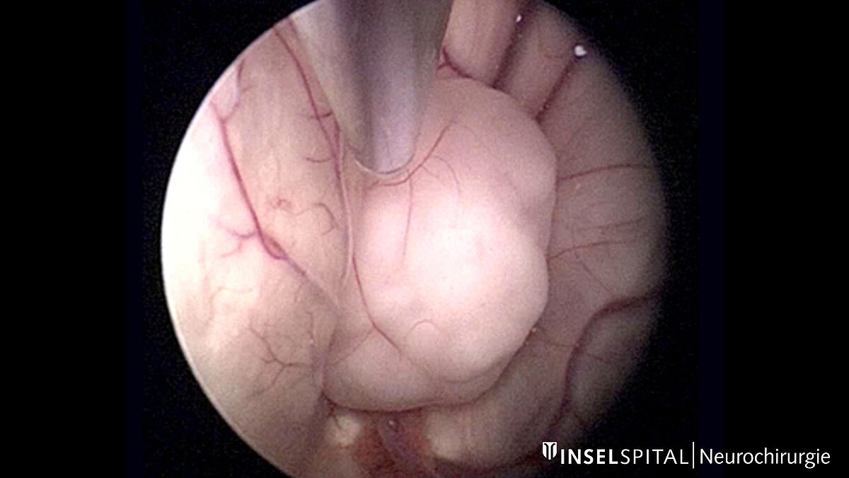
Colloid cysts are benign, cystic structures lined with an epithelial layer and filled with a rather mucinous fluid. Colloid cysts are often located in the third ventricle and, depending on their location and size, can block the pathways of cerebrospinal fluid (CSF). Very small, asymptomatic colloid cysts usually do not require surgical intervention. However, if the colloid cyst causes symptoms or if the cyst has reached a certain size, surgical removal is generally recommended *. If surgical cyst resection is necessary, it can be performed either microsurgically or, in favorable cases, purely endoscopically, depending on the location and size of the cyst.
Tumors
Endoscopic biopsy
Brain tumors are usually operated on microsurgically. An exception are intraventricular or paraventricular tumors, i.e., tumors that grow in the brain ventricles or in close proximity to them. These do not always need to be surgically removed; some of these tumor types respond well to other forms of treatment. However, a fine tissue examination of the tumor is usually necessary to diagnose and determine further therapy. If the tumor is located at an easily accessible site, an endoscopic biopsy is possible. In contrast to conventional biopsy, the endoscopic approach offers a distinct advantage due to the visual view of the tumor. Thus, the surgeon can visually see the exact biopsy site – a fact that leads to an increased diagnosis rate. Likewise, minor bleeding caused by the biopsy can be stopped directly. Because tumors in the ventricular system can obstruct CSF outflow, the neuroendoscopic approach provides the opportunity to directly perform an endoscopic third ventriculostomy or septostomy.
Full endoscopic tumor resection
In some instances, a fully endoscopic removal of an intraventricular tumor is possible. Tumors with a moderate blood vessel supply, a soft consistency, and a size of < 2–3 cm are particularly suitable for this *. However, if the tumor shows adhesions to structures such as the fornix or the thalamostriate vein, a complete resection is often not possible in order to avoid injury to these structures *. The disadvantage of neuroendoscopic tumor surgery for the surgeon primarily lies in the restricted maneuverability afforded by the endoscope and the absence of bimanual dexterity, as is possible in an open surgery.
Pituitary tumors and skull base surgery
Endoscopes enable the surgeon to obtain excellent illumination of the surgical area and correspondingly good image quality of the surgical site, even via a long and narrow access path.
Endoscopes also allow an angled view into areas that cannot be seen directly through the operating microscope. Thus, in addition to the classic transcranial approach, the endoscopic approach can also be used to perform transnasal-controlled surgery at the base of the skull. For example, pituitary tumors can be completely removed under visual control.
Surgery in the area of the brain stem and cranial nerves
In the area of the brain stem and cranial nerves, many sensitive structures are located together in a very confined space. During operations in this area, the endoscope allows a view behind these structures without damaging them. Thus the individual anatomical conditions can be recorded, and causes of disturbances can be identified. This includes, for example, a pulsating vascular loop as the cause of trigeminal neuralgia or hemifacial spasm. With the help of the endoscope, the microvascular conflict of vessel and nerve can be precisely localized and its correction visualized since neuroendoscopy allows precise visualization of the anatomical conditions at the cerebellopontine angle from multiple angles *.
-
The Principles of Neurological Surgery. Archives of Neurology And Psychiatry. 1942;48(1):162.
-
Gaab M, Schroeder H. Neuroendoskopie und endoskopische Neurochirurgie. Der Nervenarzt. 1997;68(6):459-465.
-
Cappabianca P, Cinalli G, Gangemi M, et al. Application of neuroendoscopy to intraventricular lesions. Neurosurgery. 2008 Feb;62 Suppl 2:575-97; discussion 597-8.
-
Krähenbühl A, Baldauf J, Gaab M, Schroeder H. Endoscopic temporal ventriculocisternostomy: an option for the treatment of trapped temporal horns. Journal of Neurosurgery: Pediatrics. 2013;11(5):568-574.
-
Caemaert J, Abdullah J, Calliauw L, Carton D, Dhooge C, van Coster R. Endoscopic treatment of suprasellar arachnoid cysts. Acta Neurochir (Wien). 1992;119(1-4):68-73.
-
Schroeder H, Gaab M, Niendorf W. Neuroendoscopic approach to arachnoid cysts. Journal of Neurosurgery. 1996;85(2):293-299.
-
Mathiesen, T., Grane, P., Lindgren, L. and Lindquist, C., 1997. Third ventricle colloid cysts: a consecutive 12-year series. Journal of Neurosurgery, 86(1), pp.5-12.
-
Gaab M, Schroeder H. Neuroendoscopic approach to intraventricular lesions. Journal of Neurosurgery. 1998;88(3):496-505.
-
D'Angelo V, Galarza M, Catapano D, Monte V, Bisceglia M, Carosi I. Lateral Ventricle Tumors: Surgical Strategies According to Tumor Origin and Development – A Series of 72 Cases. Operative Neurosurgery. 2005;56(suppl_1):ONS-36-ONS-45.
-
Teo C, Nakaji P, Mobbs R. Endoscope-Assisted Microvascular Decompression for Trigeminal Neuralgia:Technical Case Report. Operative Neurosurgery. 2006;59(suppl_4):ONS-E489-ONS-E490.