Navigation techniques are now well established in neurosurgery and are standard practice for brain tumor surgery as well as spinal stabilization procedures. In vascular surgery, however, they are used only in a few highly specialized centers worldwide. Computer-assisted neuronavigation serves as an electronic orientation aid during surgery. It allows preoperative MRI or CT scans to be superimposed onto the patient’s skin surface. This enables the surgeon to continuously verify their position in relation to the anatomical structures. As a result, procedures become more precise and safer.
How does vascular navigation work?
The goal of vascular navigation is to calculate a vascular pathology such as a vascular disorder or vascular malformation (for example, an aneurysm or arteriovenous malformation) and then to virtually represent it in exactly the perspective in which the surgeon is approaching that area.
Vascular navigation is much more complex than conventional navigation techniques and is only offered at a few centers. The reason for this is that often multiple imaging techniques need to be combined to calculate the three-dimensional object: CT angiography, MRI angiography, and digital subtraction angiography. Additionally, information about blood flow in the vascular disorder needs to be incorporated, especially in arteriovenous malformations.
The three-dimensional object is displayed on a screen in the operating room but can also be directly integrated into the heads-up display of the surgical microscope. The latter is referred to as augmented reality because the images from the navigation are overlaid directly onto the normal image seen through the surgical microscope.
How is vascular navigation used during surgery?
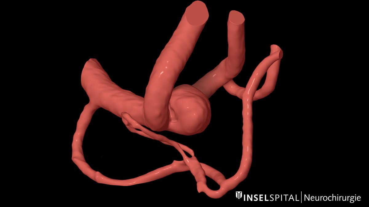
Aneurysm operation
With the help of vascular navigation, the surgeon in an aneurysm operation, for example, can precisely see at any point the position of the aneurysm, the location of the vessels branching off from it, and the risk areas on the aneurysm sac before the actual exposure of the aneurysm during surgery. The advantages of this navigation technique are a more targeted and therefore less invasive preparation, as well as reduced manipulation of the aneurysm itself *.
Without vascular navigation, the surgeon would have to expose all branching vessels of the aneurysm. Thanks to vascular navigation, the exposure can be targeted to the necessary areas. Therefore, the procedure means less invasiveness and more safety for the patient during surgery.
Bypass operation
In a bypass operation, vascular navigation assists in locating suitable donor and recipient vessels *. This enables the surgery to be performed more quickly, with less invasiveness, and with greater precision.
-
Raabe A, Beck J, Rohde S, Berkefeld J, Seifert V. Three-dimensional rotational angiography guidance for aneurysm surgery. Journal of Neurosurgery. 1. September 2006;105(3):406–11.
-
Rychen J, Goldberg J, Raabe A, Bervini D. Augmented Reality in Superficial Temporal Artery to Middle Cerebral Artery Bypass Surgery: Technical Note. Oper Neurosurg (Hagerstown). 1. April 2020;18(4):444–50.