
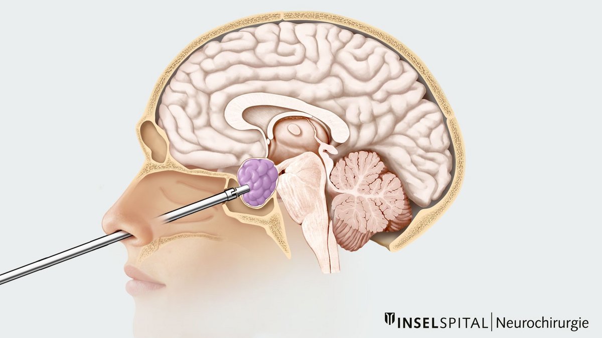
During endoscopic pituitary surgery, we remove the pituitary adenoma using a minimally invasive technique through the nose. This direct transnasal approach provides a clear view of the tumor and the surrounding structures without the need to displace the brain. Neurosurgeons and otolaryngologists work closely together to remove the tumor as safely as possible while preserving nasal function.
Why is the surgical approach through the nose?
The pituitary gland is located at the base of the skull in a bony recess known as the sella turcica. Above it are the optic nerves and the optic chiasm. The path through the nasal cavity and the sphenoid sinus behind it leads directly to the pituitary gland from below. As a result, no skin incision is required, and the brain does not need to be moved aside.
Specialists in neurosurgery and otolaryngology establish the access pathway together using an endoscope. This thin instrument, equipped with a camera and light source, transmits magnified images to a screen. Every effort is made to preserve the nasal structures and their function as much as possible.
Access to the pituitary gland
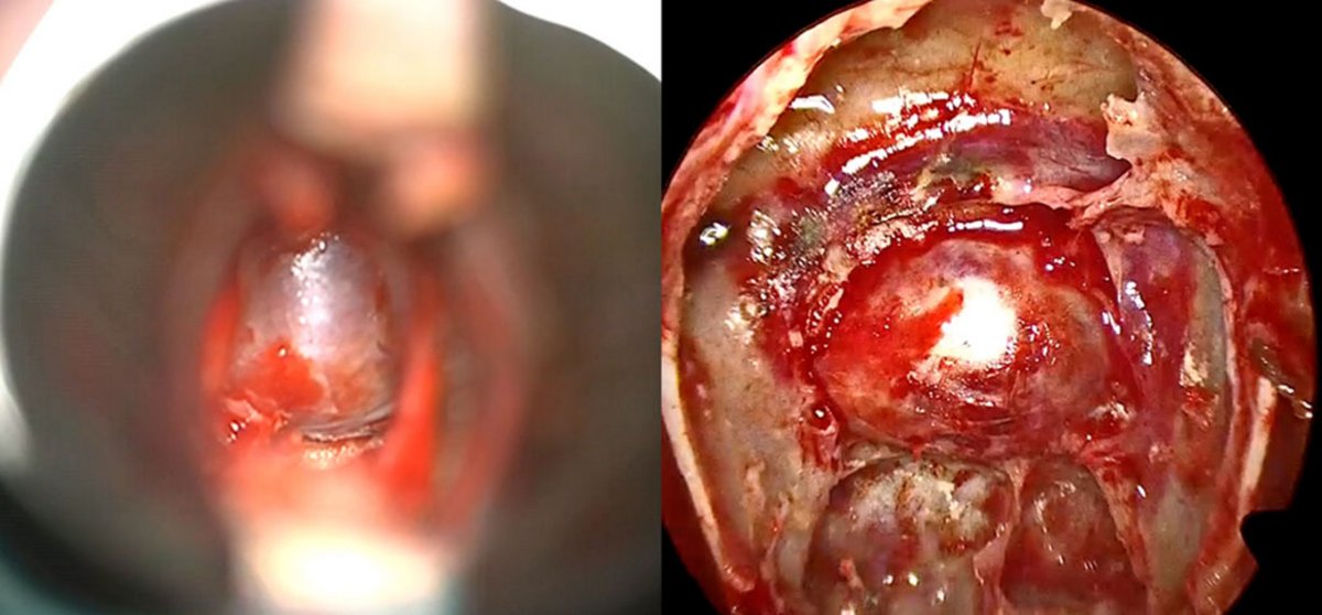
Depending on the size and extent of the tumor, as well as the individual anatomical conditions, the surgical team operates through one or both nostrils. The nasal turbinates are preserved as much as possible and temporarily moved to the side. The front wall of the sphenoid sinus is then opened. This provides access to the bony floor of the sella turcica. After opening the bone and the dura mater, the tumor and the healthy pituitary gland become visible.
Removal of the tumor
The surgical team carefully dissects the tumor margins and removes the adenoma. If the tumor’s location and size allow, it is removed in one piece whenever possible. Larger tumors are often first reduced in size from the inside and then gradually dissected out.
Skull base reconstruction
After the tumor has been removed, the surgical opening at the skull base is carefully closed.
In straightforward cases, collagen or fibrin glue patches are sufficient. If cerebrospinal fluid (CSF) leaks during the procedure, additional measures are required. Depending on the findings, the surgical team may use the patient’s own abdominal fat tissue, suture the dura mater, place a small piece of bone, or use a sugar matrix.
For larger defects or extended surgical approaches, the team covers the area with a well-vascularized mucosal flap taken from the nasal septum (nasoseptal mucosal flap). This tissue generally heals very well.
Nasal packing is not routinely required after the procedure.
What are the advantages of endoscopy?
The endoscope has a diameter of approximately 4 millimeters and provides a magnified panoramic view of the surgical field. Angled endoscopes also allow the surgical team to visualize areas located to the side and to remove tumor tissue using curved instruments.
This can offer advantages over the microscopic technique, particularly for large, invasive, or hormone-active tumors—for example, in Cushing’s disease or acromegaly *, *, *. Extended approaches to the skull base are also possible using endoscopic techniques.
For small, centrally located tumors, microscopic and endoscopic procedures can achieve comparable results. In a large study, new deficits involving hormone axes occurred less frequently after endoscopic procedures than after microscopic procedures: 9.7 % compared with 28.4 % *. One possible explanation is the improved visualization of the healthy pituitary gland.
Are there any disadvantages to the endoscopic technique?
- The duration of the operation depends greatly on the size of the tumor and the individual anatomical conditions. Studies report an average operating time of 3–5 hours *, *, *. Many uncomplicated tumors can be safely removed within 1.5–3 hours.
- The procedure generally requires two surgeons: one to operate the instruments and the other to control the endoscope.
- Thewound surface of the nasal mucosa may be somewhat larger than with the microscopic technique. Nasal-related quality of life is comparable with both procedures *. Temporary disturbances of the sense of smell occur more frequently after endoscopic procedures, but they very rarely persist permanently *.
At Inselspital, we perform pituitary surgeries entirely endoscopically.
What happens after the operation?
After the procedure, patients are monitored for at least one night in the Intermediate Care Unit (IMC). The medical team closely monitors, in particular, hormone levels and the body’s salt and water balance. If the recovery is uncomplicated, patients can usually be discharged home after 3–5 days.
Detailed information on what to expect after the operation, including recommendations for recovery and follow-up examinations, can be found in our patient information brochure (in German).
What complications might occur?
Endoscopic endonasal pituitary surgery is generally a safe procedure. However, as with any surgery, complications are possible. Your individual risk depends, among other factors, on the type, size, and extent of the tumor, as well as on your anatomical conditions. The treatment team will discuss your specific situation with you in detail before the surgery.
- Cerebrospinal fluid leak: In about 3–9% of cases, cerebrospinal fluid leaks from the nose after surgery *, *, *, *, *. This is caused by a tear in the thin membrane covering the tumor. In most cases, this tear must be repaired with another surgery.
- New hormonal imbalance: In 5–20% of cases, hormone replacement therapy with medication is necessary in the medium or long term *, *, *, *. Many hormonal imbalances are temporary.
- Temporary ADH deficiency (diabetes insipidus): In about 8–15% of cases, a temporary disturbance in water and salt balance occurs *, *. This very rarely persists long-term.
- Syndrome of inappropriate antidiuretic hormone secretion (SIADH): In about 15–30% of cases, the body temporarily retains too much water *. Limiting fluid intake as a precaution can largely prevent severe complications. In most cases, fluid balance normalizes within 2–3 weeks.
- Severe nosebleeds: about 1–8% *, *, *.
- Cranial nerve dysfunction with double vision: In uncomplicated pituitary adenomas, this occurs in less than 1 % of cases *. The risk is higher with hormone-secreting tumors that have invaded the adjacent cavernous sinus. In most cases, the symptoms resolve within several months.
Our results in the quality comparison
We continuously track our treatment outcomes and compare them with international data. The following figures were obtained for endoscopic surgeries for pituitary adenomas performed at Inselspital between April 2025 and April 2026. Malignant tumors and extended approaches to the skull base are not included in this analysis.
| Result | Percentage |
| Another surgery due to a cerebrospinal fluid leak | 3.7% |
| Another surgery for residual tumor | 3.7% |
| New hormone axis replacement therapy | 14.8%* |
| Injury to the internal carotid artery | 0% |
| Meningitis | 0% |
| Deaths | 0% |
* Long-term results are not yet available.
Our treatment principles
- Neurosurgery and otolaryngology work closely together.
- We regularly review our treatment outcomes to continuously improve quality.
- We plan each surgery on an individual basis, taking risks into account. We perform a surgical procedure only if the expected benefit justifies the associated risk.
- We place great emphasis on preserving the structure and function of the nose and carefully planning postoperative care.
We provide open and transparent information about the goal of the surgery, the prospects for success, possible risks, and treatment alternatives.
-
Dorward NL. Endocrine outcomes in endoscopic pituitary surgery: a literature review. Acta Neurochir (Wien). 2010;152(8):1275-9.
-
Tang OY, Chen JS, Monje S, Kumarapuram S, Eloy JA, Liu JK. Comparison of Surgical Modalities for Giant Pituitary Adenoma: A Systematic Review and Meta-Analysis of 1413 Patients. Oper Neurosurg. 2025;28(1):1-18.
-
Chen CJ, Ironside N, Pomeraniec IJ, Chivukula S, Buell TJ, Ding D, et al. Microsurgical versus endoscopic transsphenoidal resection for acromegaly: a systematic review of outcomes and complications. Acta Neurochir (Wien). 2017;159(11):2193-207.
-
Little AS, Kelly DF, White WL, Gardner PA, Fernandez-Miranda JC, Chicoine MR, et al. Results of a prospective multicenter controlled study comparing surgical outcomes of microscopic versus fully endoscopic transsphenoidal surgery for nonfunctioning pituitary adenomas: the Transsphenoidal Extent of Resection (TRANSSPHER) Study. J Neurosurg. 2020;132(4):1043-53.
-
Gao Y, Zhong C, Wang Y, Xu S, Guo Y, Dai C, et al. Endoscopic versus microscopic transsphenoidal pituitary adenoma surgery: a meta-analysis. World J Surg Oncol. 2014;12:94.
-
Gurses ME, Gökalp E, Gecici NN, Shah KH, Baboun SR, Eatz TA, et al. The learning curve and outcomes of 1038 endoscopic endonasal transsphenoidal pituitary tumor surgeries - A single surgical team experience. Surg Neurol Int. 2024;15:407.
-
Osborne C, Lewis D, Dixon B, Caputo C, Magee A, Gnanalingham K, et al. Equivalent outcomes in nasal symptoms following microscopic or endoscopic transsphenoidal surgery: results from multi-centre, prospective study. Acta Neurochir (Wien). 2022;164(6):1589-97.
-
Starke RM, Raper DM, Payne SC, Vance ML, Oldfield EH, Jane JA, Jr. Endoscopic vs microsurgical transsphenoidal surgery for acromegaly: outcomes in a concurrent series of patients using modern criteria for remission. J Clin Endocrinol Metab. 2013;98(8):3190-8.
-
Dallapiazza R, Bond AE, Grober Y, Louis RG, Payne SC, Oldfield EH, et al. Retrospective analysis of a concurrent series of microscopic versus endoscopic transsphenoidal surgeries for https://doi.org/10.1186/1477-7819-12-94Knosp Grades 0-2 nonfunctioning pituitary macroadenomas at a single institution. J Neurosurg. 2014;121(3):511-7.
-
Stroud A, Dhaliwal P, Alvarado R, Winder MJ, Jonker BP, Grayson JW, et al. Outcomes of pituitary surgery for Cushing's disease: a systematic review and meta-analysis. Pituitary. 2020;23(5):595-609.
-
Thakur JD, Corlin A, Mallari RJ, Yawitz S, Eisenberg A, Sivakumar W, et al. Complication avoidance protocols in endoscopic pituitary adenoma surgery: a retrospective cohort study in 514 patients. Pituitary. 2021;24(6):930-42.
-
Fatemi N, Dusick JR, Mattozo C, McArthur DL, Cohan P, Boscardin J, et al. Pituitary hormonal loss and recovery after transsphenoidal adenoma removal. Neurosurgery. 2008;63(4):709-18; discussion 18-9.
-
Agam MS, Wedemeyer MA, Wrobel B, Weiss MH, Carmichael JD, Zada G. Complications associated with microscopic and endoscopic transsphenoidal pituitary surgery: experience of 1153 consecutive cases treated at a single tertiary care pituitary center. J Neurosurg. 2019;130(5):1576-83.
-
Losa M, Mortini P, Barzaghi R, Ribotto P, Terreni MR, Marzoli SB, et al. Early results of surgery in patients with nonfunctioning pituitary adenoma and analysis of the risk of tumor recurrence. J Neurosurg. 2008;108(3):525-32.