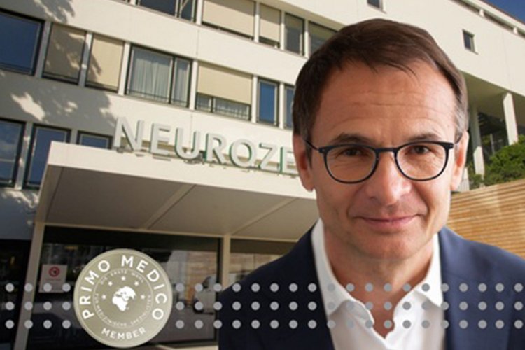
New senior physicians in neurosurgery
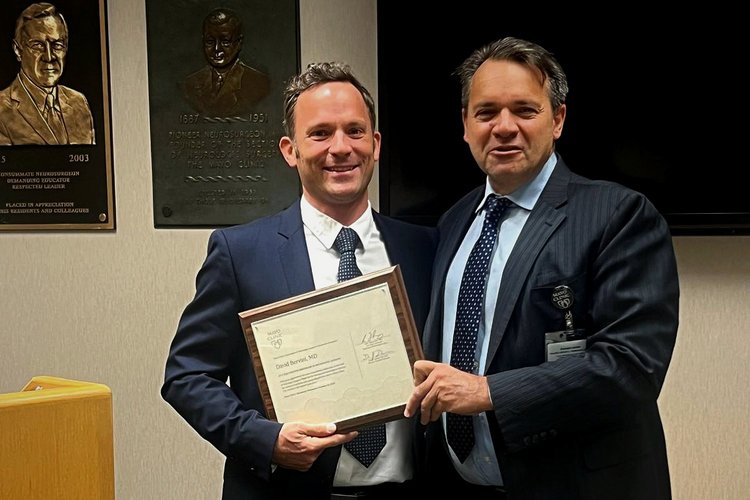
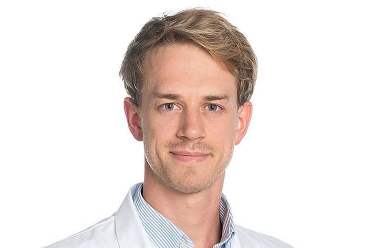
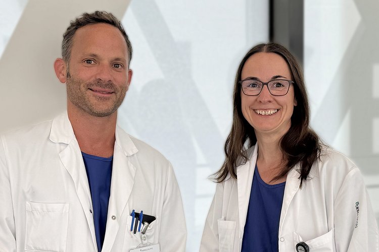
Prof. Kathleen Seidel, MD, and Prof. David Bervini, MD, will be appointed to the senior medical staff on September 1, 2025. The Department of…
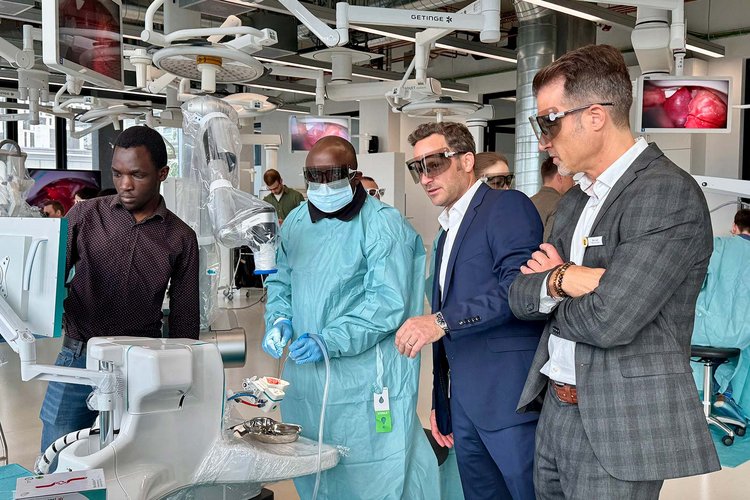
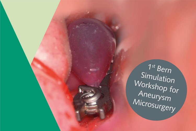
Vascular neurosurgery is a highly specialized discipline. Research in this field is crucial for continuously improving the treatment of life-threatening vascular conditions such as aneurysms, strokes, and vascular malformations, as well as for minimizing risks to patients. Through ongoing innovations, procedures can be made safer, more precise, and less invasive, ultimately improving patients’ chances of recovery and long-term quality of life.
Vascular surgery research has made enormous progress in several key areas in recent years.
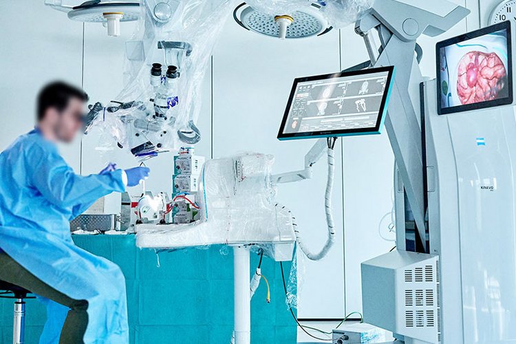
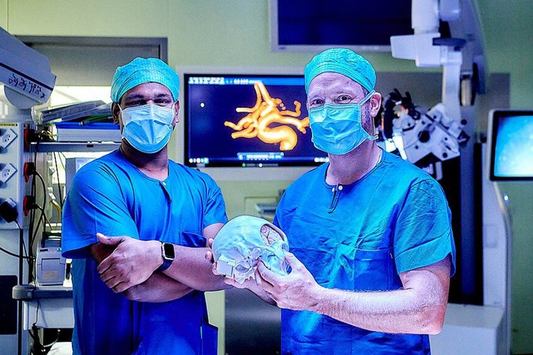
Firstly, the development of high-resolution imaging techniques such as 3D angiography and functional magnetic resonance imaging (MRI) has made the planning and execution of procedures significantly more precise.
Secondly, new minimally invasive methods, in particular endovascular techniques such as stenting and coiling, enable the treatment of brain aneurysms and vascular malformations without open surgery.
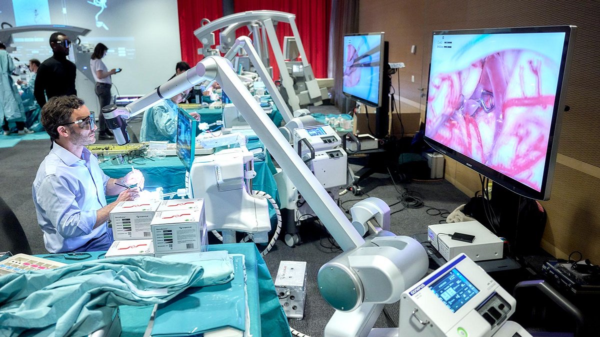
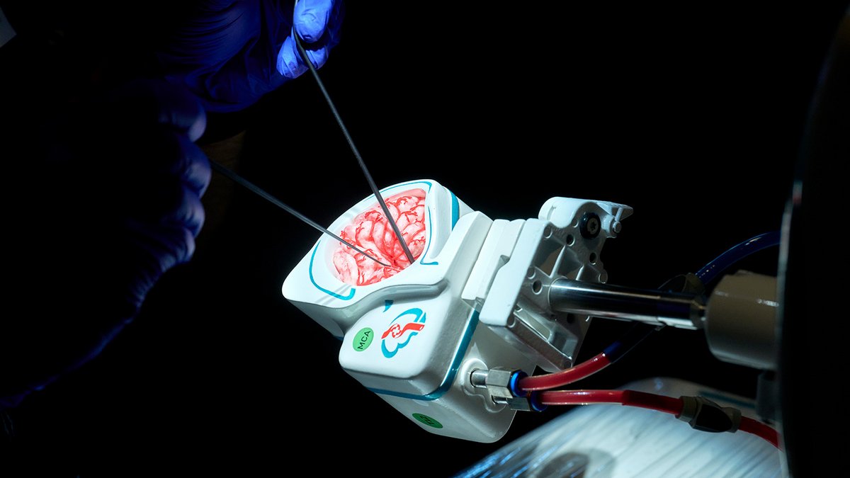
Robotics and the use of surgical microscopes with fluorescence-enhanced imaging are also improving the precision and safety of procedures.
Another advancement is the use of artificial intelligence, which helps to detect stroke and bleeding risks at an early stage and to tailor treatment plans to the individual patient.
Finally, improved biomaterials and personalized vascular implants are helping to improve the long-term prognosis and durability of procedures.
Swiss pilot screening study for unruptured intracranial aneurysms to prevent subarachnoid hemorrhage in smokers
The aim of the study is to find out whether smokers between 30 and 60 years of age who have smoked more than one pack of cigarettes per day on average for 10 years (10 pack-years) have an increased incidence of unruptured aneurysms of the cerebral vessels. Furthermore, we would like to investigate the consequences of such an examination on further treatment if an aneurysm is discovered (if necessary, elimination of the aneurysm, smoking cessation) and quality of life.
Approximately 3% of the population have a so-called aneurysm of the cerebral vessels. These aneurysms are bulges in the blood vessels that can burst and cause a life-threatening cerebral haemorrhage. As aneurysms of the cerebral vessels do not cause any symptoms, they are usually discovered by chance or as soon as a cerebral haemorrhage occurs.
Smoking is one of the most important risk factors for the development of a cerebral hemorrhage from an aneurysm. 40% of all patients with cerebral hemorrhages from aneurysms are smokers. If smokers are known to have an unruptured aneurysm, the risk of a cerebral hemorrhage later in life is increased.
Recent findings from smaller studies suggest that smokers are significantly more likely to have aneurysms of the brain vessels (> 10%) than the general population (3%). This raises the question of whether there is a benefit to screening heavier smokers, as is done for other diseases such as bowel cancer, in order to prevent future cerebral hemorrhages.
| Head of study: | PD Dr. Johannes Goldberg, MD |
| Study sponsor: | Prof. Dr. David Bervini, MD, MAdvSurg |
| Study coordinator: | Nicole Söll |
Early minimally invasive image guided endoscopic evacuation of intracerebral haemorrhage (EMINENT-ICH): a randomized controlled trial
Spontaneous cerebral hemorrhage is the second most common type of stroke, affecting approximately 2500 people in Switzerland every year. One year after a cerebral hemorrhage, around half of those affected have died, while many of the survivors are severely impaired.
Currently, there is no effective treatment for cerebral hemorrhage. The standard treatment consists of controlling blood pressure, intensive monitoring and early rehabilitation. Open brain surgery is also often performed to remove the bleeding. However, neither this nor the standard treatment shows any clear advantages for survival or quality of life for those affected.
However, minimally invasive endoscopic removal of the bleeding point could offer advantages, as smaller studies suggest. However, large, randomized studies are needed to confirm these results. The EMINENT-ICH study aims to show that minimally invasive endoscopic intervention together with standard treatment can offer better chances of survival and less permanent damage for patients.
| Head of study: | Prof. Dr. Andreas Raabe, MD |
| Study coordinator | Dr. Lisa Grönnert, PhD |
| Study identifier: | NCT04805177 |
A proteomics discovery approach to identify candidate biomarkers of atherosclerotic plaque instability in endarterectomy specimens of patients with carotid disease
An important cause of strokes is carotid stenosis, a narrowing of the carotid artery caused by deposits. The narrowing is caused by arteriosclerosis, in which fats and cholesterol crystals are deposited in the artery walls. If these deposits tear, small particles can be released, which enter the brain via the bloodstream and trigger a stroke. It is hypothesized that certain compositions of the deposits are associated with a higher risk of tearing and thus of stroke.
In our research project, we are investigating the exact composition of these deposits and comparing them between different patient groups. We hope this will enable us to identify deposits with an increased risk of rupture at an early stage so that targeted treatment measures can be taken to prevent strokes.
| Head of study: | Prof. Dr. Andreas Raabe, MD |
| Study coordinator: | Dr. Lisa Grönnert, PhD |

This study looks at whether combining routine medical imaging with computational fluid dynamics (CFD) – a method to analyze blood flow – can help identify patients at higher risk of stroke and improve treatment planning.
A narrowing of the carotid artery (carotid stenosis, CAS) affects about 3–6% of people over the age of 70 and becomes more common with age. It is responsible for roughly one in five ischemic strokes or transient ischemic attacks (TIA). The greatest danger arises when a plaque in the artery wall ruptures, leading to a blood clot that blocks blood flow to the brain. Since about one quarter of patients with symptom-free CAS already have such «vulnerable plaques», early detection is important to select those who could benefit from surgery (carotid endarterectomy) to prevent stroke.
In this study, we aim to test whether CFD models, based on data already collected in routine care, can be used to better select patients for treatment compared with current imaging standards. In the long term, this approach could also benefit patients in regions with limited medical resources.
Heads of study: Prof. David Bervini, MD and Dr. Shaokai Zheng
Study on the follow-up of unruptured intracranial aneurysms using 7-Tesla high-field MRI
Approximately 3% of the population has a cerebral aneurysm – a bulge in a blood vessel in the brain that, in rare cases, can rupture and cause a life-threatening brain hemorrhage. For those affected, an accurate risk assessment is crucial in order to decide whether treatment is necessary or regular monitoring is sufficient.
Why is a more detailed examination important?
Aneurysms with an increased risk of rupture often show inflammation in the vessel wall. This can be made visible using contrast agents. However, the resolution of a conventional 3T MRI is often insufficient to reliably detect this inflammation.
7T high-field MRI – a new dimension in imaging
A 7T high-field MRI offers significantly higher image resolution than a standard 3T MRI. This allows the condition of the aneurysm wall to be assessed more accurately and the individual risk of rupture to be better estimated.
Opportunity to participate in this study
As part of our research project, we would like to perform a high-resolution 7T high-field MRI scan on patients with unruptured aneurysms in addition to the regular follow-up examinations using 3T MRI. Study participants will thus make a valuable contribution to improving diagnostics and developing even safer treatment strategies.
| Head of study: | PD Dr. Johannes Goldberg, MD |
| Study sponsor: | Prof. Dr. David Bervini, MD, MAdvSurg |
| Study coordinator: | Nicole Söll |