
In certain hormone-active pituitary adenomas, tumor cells can invade the wall of an adjacent venous sinus, the cavernous sinus. Using modern endoscopic surgical techniques, this wall can be removed in select cases to increase the chance of complete tumor removal and hormonal cure. The treatment team decides on a case-by-case basis whether this additional surgical step is appropriate, after carefully weighing the benefits and risks.
What is the cavernous sinus?
The cavernous sinus is a large venous sinus located directly next to the pituitary gland. Important cranial nerves and the internal carotid artery (arteria carotis interna) pass through it. The pituitary gland drains some of its venous blood through small natural openings in the inner wall of the cavernous sinus. Cells from a pituitary adenoma can also enter the wall or the cavernous sinus through these openings. How often this occurs depends, among other things, on the type of tumor *.
In the past, the cavernous sinus was generally not opened during pituitary surgery; as a result, tumor fragments in this area were often left behind. Today, modern endoscopes and specialized instruments allow for the targeted removal of these tumor fragments and the affected wall in selected patients.
When is this surgical technique an option?
Whether the medial wall of the cavernous sinus should be removed depends on various factors:
- Type of tumor
- Extent of infiltration
- Patient's age
- Treatment alternatives
- Individual surgical risk
It is particularly important to distinguish between non-functioning pituitary adenomas and hormonally active pituitary adenomas.
Non-functioning pituitary adenomas
In the case of non-functioning adenomas, removal of the wall is usually not warranted, especially in older patients. A small residual tumor can often be monitored with regular checkups or, if necessary, treated with radiation therapy.
Functioning pituitary adenomas
In cases of functioning adenomas ––such as those associated with Cushing’s disease or acromegaly– it is particularly important to remove the tumor as completely as possible. Even a small number of remaining tumor cells in the cavernous sinus or its wall can lead to continued excessive hormone production.
In carefully selected cases, removal of the medial wall can therefore improve the prognosis for hormonal remission *, *, *, *. This challenging technique should only be performed at centers with appropriate experience.
What do studies show?
Recent studies report remission rates of 84–93% *, *, * for hormone-secreting pituitary adenomas following the use of this technique. The remission rate indicates the proportion of treated patients in whom the disease is no longer detectable or has significantly regressed following therapy.
Tumor cells were detected in the resected wall segments in 70–80% of cases *, *, *.
Even when no tumor invasion was visible during surgery, tumor cells were nevertheless found in this wall under the microscope in 57% of cases *.
These results underscore that the medial wall is a potential site for residual tumor tissue in hormone-active adenomas.
How is the medial wall removed?
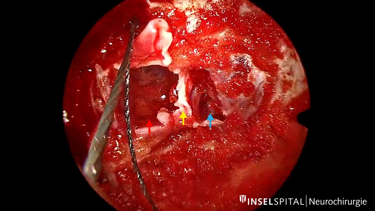
The surgery is performed endoscopically through the nose and is part of the procedure to remove the pituitary adenoma. After the main portion of the tumor has been removed, the surgical team proceeds step by step:
- Incision: First, the anterior wall of the cavernous sinus is carefully incised.
- Hemostasis: Since this is a venous blood vessel, the area is filled with a special foam paste and temporarily packed to control bleeding.
- Detachment of the wall: The medial wall is gradually incised at its lower and upper attachment points and detached from the surrounding structures.
- Removal: The detached portion of the wall can then be removed along with any tumor tissue and examined histologically.
What are the risks?
The internal carotid artery and important cranial nerves run through the cavernous sinus. Therefore, injury to these structures cannot be ruled out. The individual risk depends on the anatomy and the extent of the tumor and will be discussed in detail before surgery.
- Injury to the internal carotid artery: This complication can be serious. Data published to date indicate a very low rate among experienced surgical teams.
- Temporary cranial nerve palsy: This may manifest primarily as double vision and occurred in 7.1 % of patients in one study *. Function usually recovers completely, but recovery may take several months.
Our approach at Inselspital
At Inselspital, we offer resection of the medial wall of the cavernous sinus for selected patients. This procedure can improve treatment outcomes for hormone-secreting pituitary adenomas, but it is not suitable for every situation.
We recommend this additional surgical step only if the expected benefit outweighs the individual risk, and we discuss the basis for this decision in detail with the patient before the procedure.
-
Mohyeldin A, Katznelson LJ, Hoffman AR, Asmaro K, Ahmadian SS, Eltobgy MM, Nayak JV, Patel ZM, Hwang PH, Fernandez-Miranda JC. Prospective intraoperative and histologic evaluation of cavernous sinus medial wall invasion by pituitary adenomas and its implications for acromegaly remission outcomes. Sci Rep. 2022 Jun 15;12(1):9919. doi: 10.1038/s41598-022-12980-1.
-
Oldfield EH. Cushing's Disease: Lessons Learned From 1500 Cases. Neurosurgery. 2017 Sep 1;64(CN_suppl_1):27-36. doi: 10.1093/neuros/nyx378.
-
Cohen-Cohen S, Gardner PA, Alves-Belo JT, Truong HQ, Snyderman CH, Wang EW, Fernandez-Miranda JC. The medial wall of the cavernous sinus. Part 2: Selective medial wall resection in 50 pituitary adenoma patients. J Neurosurg. 2018 Sep 7;131(1):131-140. doi: 10.3171/2018.5.JNS18595.
-
Constanzo F, Rychen J, Lee CK, Decker JH, Fischbein N, Johnstone T, Ljubimov V, Vigo V, Fernandez-Miranda JC. Patterns of invasion of the medial wall of the cavernous sinus by pituitary adenomas. J Neurosurg. 2025 Jun 27;143(4):895-906. doi: 10.3171/2025.3.JNS242823.
-
Truong HQ, Lieber S, Najera E, Alves-Belo JT, Gardner PA, Fernandez-Miranda JC. The medial wall of the cavernous sinus. Part 1: Surgical anatomy, ligaments, and surgical technique for its mobilization and/or resection. J Neurosurg. 2019 Jul 1;131(1):122-130. doi: 10.3171/2018.3.JNS18596. Epub 2018 Sep 7.
-
Nagata Y, Takeuchi K, Yamamoto T, Ishikawa T, Kawabata T, Shimoyama Y, Wakabayashi T. Removal of the Medial Wall of the Cavernous Sinus for Functional Pituitary Adenomas: A Technical Report and Pathologic Significance. World Neurosurg. 2019 Jun;126:53-58. doi: 10.1016/j.wneu.2019.02.134. Epub 2019 Mar 5.
-
Nishioka H, Fukuhara N, Horiguchi K, Yamada S. Aggressive transsphenoidal resection of tumors invading the cavernous sinus in patients with acromegaly: predictive factors, strategies, and outcomes. J Neurosurg. 2014 Sep;121(3):505-10. doi: 10.3171/2014.3.JNS132214. Epub 2014 Jul 11.
-
Torres A, Muñoz F, Sanmillan JL, Gabarros A. Cavernous Sinus Medial Wall Resection in Invasive Pituitary Adenomas: Outcome in Acromegaly. World Neurosurg. 2026 May;209:124929. doi: 10.1016/j.wneu.2026.124929. Epub 2026 Mar 19.