
Tethered cord syndrome, or TCS for short, is a rare condition in which the spinal cord is abnormally fused to the lower part of the spine, severely limiting normal growth. TCS can be diagnosed prenatally, shortly after birth, or during childhood. Symptoms are varied and include back pain, weakness in the legs, and problems with bladder and bowel function. Treatment often requires surgery to release the fixation in order to preserve spinal cord function and prevent further damage. Early diagnosis and intervention are crucial here to ensure the child’s optimal development and health.

How does tethered cord syndrome develop?
Generally, TCS is classified into a congenital (primary) form and an acquired (secondary) form.
Primary tethered cord syndrome
The development of the central nervous system is highly complex. As early as the first weeks of embryonic development, the formation of the spinal cord begins with the closure of the neural tube *. Abnormalities during these developmental stages can lead to permanent malformations, which are collectively referred to as dysraphia. These neural tube defects can cause a wide range of symptoms and problems.
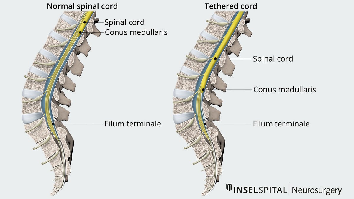
Normally, the spinal cord is largely mobile within the spine and is fixed only by the filum terminale, a delicate cord at the lower end of the spinal canal. This ensures the necessary mobility of the spinal cord during movement. However, due to various causes, the filum terminale may become «fatty», thickened, or otherwise altered, causing it to fuse with the meninges surrounding the spinal cord. In such cases, the spinal cord is no longer freely mobile. This results in fixation or even a low-lying position of the spinal cord. Movements can then lead to overstretching and, consequently, damage to the spinal cord.
Tethered cord syndrome can also occur as a result of other conditions, including myelomeningoceles (MMC), lipomyelomeningoceles, and split cord malformations *. In such cases, the spinal cord or nerve roots also become adherent to the meninges. TCS occurs more frequently, particularly in children with syndromic disorders such as VACTERL association *.
Secondary tethered cord syndrome
In much rarer cases, TCS occurs secondarily, that is, it is «acquired». This can occur, for example, following trauma, infections, or surgery in the lumbar spine.
What are the symptoms of tethered cord syndrome?
Diagnosing TCS in infants and children is often difficult because some symptoms can develop gradually. The most common signs – such as back pain, unsteady gait, and bladder problems – are often initially attributed to other causes during childhood.
The age-related variability of symptoms further complicates the diagnosis: While older children and adults mainly suffer from back and leg pain as well as weakness in the lower extremities, infants may exhibit delays in motor development, such as delayed walking.
Neurocutaneous signs such as dark or red spots, tufts of hair, or soft nodules in the lower back may also indicate dysraphia.
In some cases, associated malformations such as leg length discrepancies, foot deformities, and scoliosis occur in combination with TCS.
In summary, TCS can present with a wide range of symptoms:
- Back pain
- Leg pain
- Numbness or tingling in the legs
- Weakness in the legs
- Atrophy of the leg muscles
- Deterioration of gait
- Urinary incontinence or urinary retention
- Recurrent urinary tract infections
- Fecal incontinence
- Paraplegic symptoms
Because of the potential effects of TCS on various organ systems, specialists from fields such as neuropediatrics, pediatric neurosurgery, pediatrics, pediatric surgery, pediatric urology, and pediatric orthopedics work closely together. This enables targeted diagnosis and the development of a personalized treatment plan. This interdisciplinary collaboration also extends through the follow-up care period.
How is tethered cord syndrome diagnosed?
In most cases, children with suspected TCS first come into contact with pediatricians and neuropediatricians, who then initiate the necessary examinations in consultation with other specialties.
For children under 3 months of age, an ultrasound examination is often performed first to obtain initial findings.
If TCS is suspected, a diagnostic magnetic resonance imaging (MRI) scan of the entire neural axis – from the head to the coccyx – is necessary to confirm the diagnosis and rule out other causes.
In specific cases where the findings are unclear, an MRI in the prone position may also be performed to assess the mobility of the spinal cord and determine whether the filum terminale is fixed *. This examination is performed by our neuroradiology specialists at Inselspital.
How is tethered cord syndrome treated?
Once the diagnosis has been made, a microsurgical procedure is planned.
The surgery is a routine procedure in neurosurgery and is performed minimally invasively. Using a surgical microscope, a small opening (fenestration) is created through the bony structures, and the dura mater is incised. The filum terminale is localized under the microscope and reliably identified using intraoperative neuromonitoring (IOM) before it is transected. This step is referred to by medical professionals as detethering.
For other causes of tethered cord syndromeTCS, such as meningoceles or spinal lipomas, the surgery is generally more complex, as it can be difficult to achieve sufficient removal of the adhesions from the spinal cord.
The risk of re-tethering – that is, the filum terminale reattaching after a successful surgery – ranges from 5.2 to 8.6% *, *. In such cases, repeat surgery may be necessary. For treated myelomeningoceles, the re-tethering rate is even slightly higher due to the complexity of this condition *.
What happens after the surgery?
After a brief period of bed rest, early mobilization can begin immediately after surgery under the guidance of a physical therapist. As a rule, there are no significant limitations following the procedure.
Follow-up care after treatment for a TCS is provided by the treating interdisciplinary medical team in collaboration with outpatient or inpatient physical therapy and/or occupational therapy.
What happens if tethered cord syndrome is left untreated?
If TCS is left untreated, it can lead over time to a worsening of existing symptoms and the appearance of new symptoms. Especially in children, whose spines are still growing, it is important to conduct regular follow-up examinations to detect such developments early on.
However, surgery generally remains the only treatment option to stop the progression of symptoms.
-
Rohen Johannes W, Lütjen-Drecoll Elke. Funktionelle Embryologie. Thieme; 6. Aufl. 2022. 204 p.
-
Greenberg Mark S. Handbook of Neurosurgery. Thieme Medical Publishers; 9th ed. 2019. 1781 p.
-
Kuo MF, Tsai Y, Hsu WM, Chen RS, Tu YK, Wang HS. Tethered spinal cord and VACTERL association. J Neurosurg. 2007 Mar;106(3 Suppl):201-4. doi: 10.3171/ped.2007.106.3.201.
-
Stamates MM, Frim DM, Yang CW, Katzman GL, Ali S. Magnetic resonance imaging in the prone position and the diagnosis of tethered spinal cord. J Neurosurg Pediatr. 2018 Jan;21(1):4-10. doi: 10.3171/2017.3.PEDS16596.
-
Finger T, Schaumann A, Grillet F, Schulz M, Thomale UW. Retethering after transection of a tight filum terminale, postoperative MRI may help to identify patients at risk. Childs Nerv Syst. 2020 Jul;36(7):1499-1506. doi: 10.1007/s00381-019-04458-9.
-
Yong RL, Habrock-Bach T, Vaughan M, Kestle JR, Steinbok P. Symptomatic retethering of the spinal cord after section of a tight filum terminale. Neurosurgery. 2011 Jun;68(6):1594-601; discussion 1601-2. doi: 10.1227/NEU.0b013e31821246c6.
-
Ferreira Furtado LM, Da Costa Val Filho JA, Dantas F, Moura de Sousa C. Tethered Cord Syndrome After Myelomeningocele Repair: A Literature Update. Cureus. 2020 Oct 14;12(10):e10949. doi: 10.7759/cureus.10949.