
Cushing’s disease is usually caused by a benign tumor of the pituitary gland, which triggers excessive production of the stress hormone cortisol. If left untreated, the disease can have serious health consequences. In many cases, surgical removal of the tumor can lead to a cure. At our specialized center, we provide patients with comprehensive, multidisciplinary care – with the goal of achieving the best possible treatment outcome.
How common is Cushing's disease?
Cushing's disease is a rare condition: Each year, about 1–2 out of every 1 million people are diagnosed with it *. Women are affected about four times more often than men *, *. The disease usually develops between the ages of 30 and 40 *, *.
What symptoms does Cushing's disease cause?
In Cushing's disease, an excess of the hormone cortisol leads to various physical and psychological symptoms. Some of these are characteristic of the disease, while others may also occur in other conditions *, *, *:
Typical physical signs include:
- Weight gain, especially around the torso, while the arms and legs remain comparatively slender
- Round, often flushed face («moon face»)
- Fat deposits in the neck area («bull neck»)
- Wide, reddish-purple stretch marks on the skin
- Thin, sensitive skin and frequent bruising
- Muscle weakness, especially in the upper arms and thighs
- New or recurring acne
- Excessive facial and body hair in women
Other possible symptoms and complications include:
- High blood pressure
- Elevated blood sugar or diabetes mellitus
- Reduced bone density and bone fractures even with minimal stress and at a younger age (< 50 years)
- Menstrual irregularities in women
- Increased susceptibility to infections
- Psychological changes such as depressive mood, anxiety, or irritability

Symptoms usually develop gradually and are sometimes nonspecific. For this reason, Cushing's disease is often not diagnosed until a late stage. A thorough hormonal evaluation is essential for making a reliable diagnosis.
What is the difference between Cushing's disease and Cushing's syndrome?
Cushing's syndrome is the umbrella term for symptoms and physical changes caused by a prolonged excess of the hormone cortisol—regardless of the cause.
In Cushing's disease, the excess cortisol is caused by a pituitary adenoma, which is usually benign. The tumor produces elevated levels of the hormone ACTH, thereby stimulating the adrenal glands to release too much cortisol. Cushing’s disease is thus a specific form of Cushing’s syndrome and, at about 60–70 %, its most common endogenous cause *, *, *.
How is Cushing's disease diagnosed?
Diagnosing Cushing's disease is a complex process that involves several steps.
First, various hormone tests are used to determine whether the body is actually producing too much cortisol. If this condition, known as hypercortisolism, is confirmed, Cushing’s syndrome is present.
Next, the cause of the excess cortisol is investigated to determine whether an ACTH-producing pituitary adenoma – and thus Cushing’s disease – is present.
Detection of excess cortisol
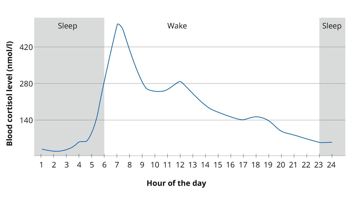
There are three tests available to detect excess cortisol. Depending on the situation, they may be combined or repeated *:
- Nighttime salivary cortisol: This test shows whether cortisol levels drop as expected at night. Shift work, stress, or smoking can influence the results *.
- 24-hour urine: This test measures total cortisol excretion over a 24-hour period. Multiple measurements are usually required.
- 1-mg dexamethasone suppression test: After taking dexamethasone in the evening, the cortisol level in the blood is measured the next morning. If it does not drop sufficiently, this may indicate excess cortisol. Medications containing estrogen can skew the results *.
The results are always evaluated in conjunction with the patient’s symptoms and other findings.
In cyclic Cushing’s disease, phases of elevated and normal cortisol levels alternate. Therefore, repeated testing may be necessary. *, *, *
Determining the cause
If excess cortisol is confirmed, the ACTH level in the blood is measured. ACTH stimulates the adrenal glands to produce cortisol.
- Low ACTH level: The cause is likely to be in one of the adrenal glands.
- Normal or elevated ACTH level: A possible cause is an ACTH-producing tumor—usually a pituitary adenoma (Cushing’s disease), or, less commonly, a tumor in another part of the body.
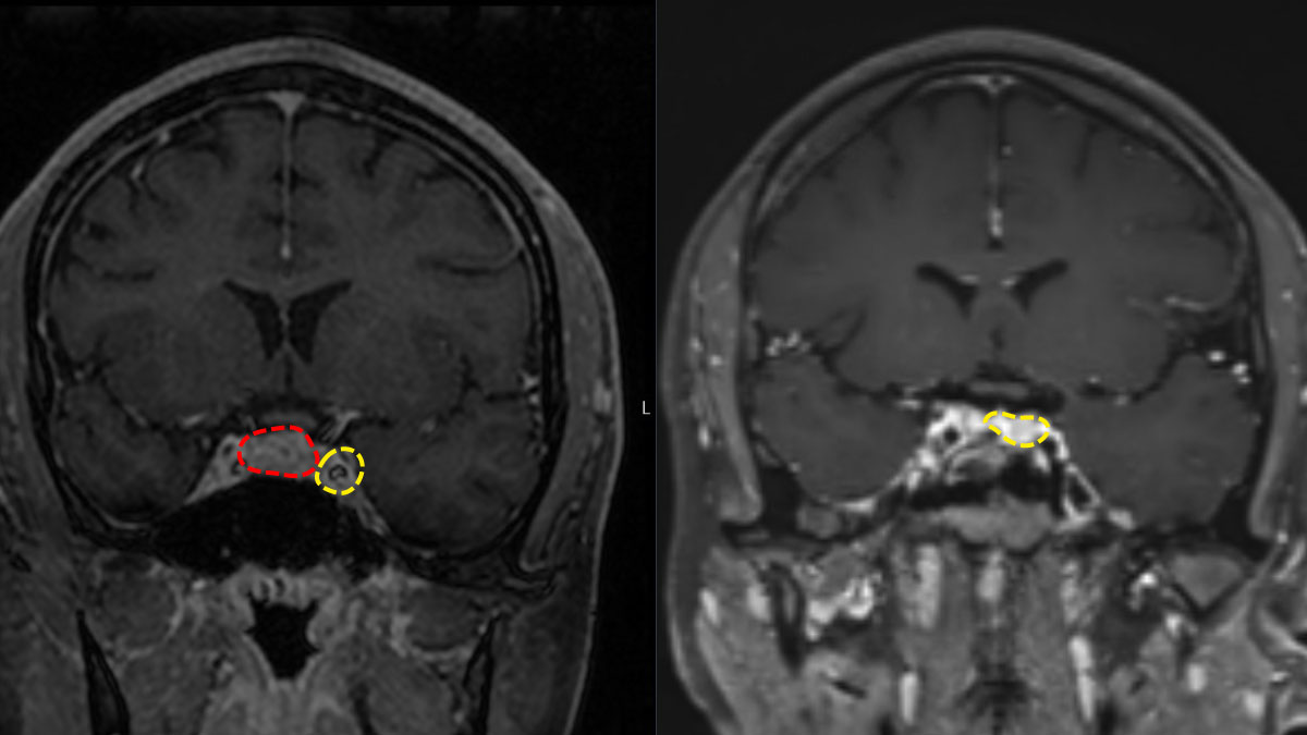
If Cushing’s disease is suspected, the pituitary gland is examined using magnetic resonance imaging (MRI). Since the tumors are often very small, special MRI sequences are used. If necessary, a high-resolution 7-tesla MRI scanner is also available at Inselspital.
Important: A pituitary adenoma visible on an MRI is not necessarily the cause of excess cortisol. Small, hormonally inactive adenomas are relatively common and are sometimes discovered incidentally. Therefore, MRI images must always be evaluated in conjunction with hormone levels.
Further tests in cases of inconclusive results
If the MRI does not reveal a clear tumor or if the cause remains unclear, additional tests may be necessary:
- Petrosal sinus sampling: ACTH levels are measured in blood vessels near the pituitary gland using a catheter. This helps determine whether the excess ACTH originates from the pituitary gland or from a tumor elsewhere in the body.
- PET/CT or other imaging techniques: These are used to search for an ACTH-producing tumor outside the pituitary gland.
- Special hormone tests: The high-dose dexamethasone suppression test, as well as desmopressin or CRH stimulation tests, can provide additional clues to the cause in selected cases.
Since the diagnostic process is complex, the findings should be evaluated by a specialized endocrinology center.
How is Cushing's disease treated?
The first-line treatment is usually surgical removal of the pituitary adenoma.
For large tumors, or when surgery alone is not sufficient, several treatment methods may be combined: Options include medications to lower cortisol levels, radiation therapy, or a combination of various approaches. In rare cases, removal of both adrenal glands may be necessary.
Surgery
Surgery is the treatment of choice. The pituitary adenoma is usually removed through the nose. In approximately 75–80 % of cases, cortisol production returns to normal after the procedure *, *, *. Since the tumors are often very small and located close to healthy pituitary tissue, extensive surgical experience and careful surgical planning are particularly important.
Depending on the tumor’s location and size, the procedure is performed endoscopically or microscopically. For larger tumors, the endoscopic technique may offer advantages; for small, centrally located adenomas, both procedures yield comparable results *, *. Whenever possible, the tumor is removed completely and along its natural borders *, *.
If the tumor is not visible on MRI
Even if the tumor is not visible, surgery may still be an option provided that the tests clearly indicate the pituitary gland as the cause. Often, petrosal sinus sampling is performed beforehand. If no adenoma is found during surgery, part of the pituitary gland may be removed in selected cases *. The chances of a cure are then approximately 60–70 %.
If the tumor cannot be completely removed
Partial removal may also be beneficial: it reduces the size of the tumor and can facilitate further treatment. Afterward, the disease can often be better controlled with medication and/or radiation therapy.
What factors influence the success of the surgery?
The success of the surgery depends on several factors:
- Experience of the surgical team: Surgeries on the pituitary gland are complex and should be performed at a specialized center. Modern endoscopic techniques allow for precise visualization of the tumor. In select cases, the adjacent wall of the cavernous sinus may also be removed *.
- Tumor size: Small tumors under 1 cm (microadénomas) can often be completely removed more frequently than larger tumors (macroadénomas) *, *.
- Visibility on MRI: A clearly identifiable tumor facilitates surgical planning.
- Location and extent: If the tumor has invaded the cavernous sinus, complete removal may be more difficult *, *.
For microadenomas (< 1 cm), cortisol production returns to normal after surgery in approximately 80–85% of cases; for macroadenomas (> 1 cm), in approximately 60–70% *, *, *.
Tissue examination after surgery
Examination of the removed tissue reveals whether the tumor was actually responsible for the hormone secretion. Certain characteristics can also provide clues about the tumor’s behavior and the risk of a relapse (recurrence).
Pituitary adenomas, also known as PitNET, are classified into different cell lines based on specific transcription factors. Tumors associated with Cushing’s disease belong to the so-called T-Pit cell line. Three types are distinguished:
- Densely granular tumors: These often cause severe Cushing’s disease, even when they are still very small.
- Loosely granular tumors: These are usually somewhat larger and often cause less distinct symptoms.
- Crook-cell adenomas: These are rare and may grow more aggressively.
Crook-cell adenomas and tumors with an elevated cell division rate are considered potentially more aggressive. In these cases, particularly frequent monitoring is recommended *, *.
It is important to note that not every tumor in the T-Pit cell lineage actually produces hormones.
Hormone levels after surgery
If the hormone-secreting pituitary adenoma has been successfully removed, the cortisol level usually drops significantly within 48 hours. For this reason, it is monitored closely during the first few days after surgery *.
A cortisol level below 55 nmol/l is a very good indication of successful treatment. In over 90 % of patients, a long-term cure can then be expected *, * , *.
A level below 140 nmol/l immediately after surgery also suggests successful treatment. However, with levels between 55 and 140 nmol/l, the risk of relapse is about 20% *.
After a successful surgery, a temporary cortisol deficiency often occurs. For this reason, cortisol is initially replaced with medication, and the dose is gradually reduced. The body’s own production usually recovers within a few months, but it may also take a year or longer. In some cases, it does not recover completely *, *.
What happens next if the surgery was not effective enough?
If disease activity persists after surgery, further treatment depends on the individual situation. If residual tumor is visible on an MRI, reoperation may be considered. This is usually more complex than the initial surgery and has a lower chance of cure. Depending on the extent of the residual tumor, a cure can be achieved in about 50–60 % of cases *, *, *.
If repeat surgery is not possible or too risky, medication or radiation therapy may be considered. Medication can control cortisol production, while radiation therapy can be used, particularly when residual tumor tissue is visible. However, the effects of radiation therapy take time to become apparent, so medication is often necessary initially *, *.
If the excess cortisol is severe after removal of the pituitary adenoma and cannot be adequately treated with medication, removal of both adrenal glands (bilateral adrenalectomy) should be considered.
Drug therapy
Various active ingredients are available for medical treatment:
- Inhibition of cortisol production: Ketoconazole, metyrapone, mitotane, and osilodrostat slow cortisol production in the adrenal glands. They do not act directly on the pituitary tumor.
- Inhibition of ACTH production: Pasireotide and cabergoline act directly on the ACTH-producing pituitary tumor.
- Blocking the effects of cortisol: Mifepristone blocks the effects of cortisol in the body.
The choice of medication depends, among other factors, on the extent of the excess cortisol, the presence of any residual tumor, and the patient’s individual comorbidities. In cases of mild excess cortisol without a visible residual tumor, cortisol-inhibiting medications are often used. If cortisol levels are significantly elevated, a combination of several active ingredients may be necessary.
If a residual tumor is visible after surgery, pasireotide or cabergoline may be considered. They reduce ACTH production and, in some cases, can also influence tumor growth *.
Radiation therapy
Radiation therapy may be considered if a residual tumor remains after surgery and the disease is not adequately controlled. It is used in particular when repeat surgery is not possible or involves high risks.
In radiosurgery, the tumor is targeted with highly precise radiation. However, it may take some time for the treatment to take full effect. During this time, medication is usually necessary to control the excess cortisol.
In approximately 50–70 % of patients treated, the disease can be controlled with radiation therapy *. If the tumor is located close to the optic nerves, treatment may be more difficult. In addition, other hormonal functions of the pituitary gland may be impaired. The therapy should be planned and carried out at a specialized center.
Removal of both adrenal glands
In cases of severe cortisol excess that cannot be adequately controlled by other methods, removal of both adrenal glands (bilateral adrenalectomy) may be considered in rare cases. This immediately stops the excess cortisol production. Subsequently, the missing adrenal hormones must be replaced with medication for the rest of the patient’s life.
In approximately 20–40% of those affected, the pituitary tumor may continue to grow afterward. This condition, known as Nelson’s syndrome, requires regular hormone and MRI monitoring *, *. If there is significant tumor growth, surgical removal is the preferred treatment whenever possible *.
What improvements can be expected after successful treatment?
After successful treatment, metabolism usually improves significantly. Body fat often decreases, even though some excess weight may remain. Existing diabetes and heart function may also improve. However, heart function does not always return to normal. In the long term, life expectancy returns to levels similar to those of the general population *.
Long-term follow-up examinations
Even after successful treatment, lifelong, regular checkups are necessary. In about 10–30% of those affected, the disease can return years later *, *, *, *, *.
Nighttime measurement of cortisol in saliva is particularly suitable for the early detection of a relapse. Once the body’s own cortisol production has recovered, this test should generally be performed annually *. In contrast, a 24-hour urine test may not indicate a relapse until later.
Why is comprehensive care important?
Cushing’s disease can affect various organ systems and lead to serious complications. Therefore, these complications must be detected early, monitored regularly, and treated specifically. These include:
- High blood pressure
- Cardiovascular diseases
- Diabetes mellitus
- Osteoporosis
- Thrombosis prevention in high-risk patients
- Infection prevention in cases of very high cortisol levels
Even if, for example, blood sugar control and heart function improve after successful treatment, regular follow-up care remains important.
When using drug therapy, potential effects on other organs must be monitored. For example, treatment with ketoconazole requires regular monitoring of liver function tests.
Why you should seek treatment at Inselspital
The Pituitary Center at Inselspital offers:
- Interdisciplinary care: Neurosurgery, ENT, endocrinology, and other specialties work closely together.
- Specialized expertise: Experienced teams treat Cushing’s disease using modern methods.
- Personalized therapy: We select a treatment plan tailored to each patient’s risk profile.
- Optimal surgical conditions: For invasive tumors, even hard-to-reach tumor portions—such as those on the wall of the cavernous sinus – can be removed endoscopically.
- Focus on cure: The goal is to remove the tumor as completely as possible and achieve long-term normalization of cortisol levels.
- Preservation of nasal function: During surgery, we take special care to minimize damage to the nose and preserve its function.
- Shared decision-making: We provide comprehensive information about the benefits and risks and determine the treatment plan together with our patients.
- Quality assurance: Structured follow-up visits and consistent quality control are an integral part of the treatment.
-
Hakami OA, Ahmed S, Karavitaki N. Epidemiology and mortality of Cushing's syndrome. Best Pract Res Clin Endocrinol Metab. 2021;35(1):101521.
-
Fleseriu M, Varlamov EV, Hinojosa-Amaya JM, Langlois F, Melmed S. An individualized approach to the management of Cushing disease. Nat Rev Endocrinol. 2023;19(10):581-99.
-
Gadelha M, Gatto F, Wildemberg LE, Fleseriu M. Cushing's syndrome. Lancet. 2023;402(10418):2237-52.
-
Guaraldi F, Zoli M, Asioli S, Corona G, Gori D, Friso F, et al. Results and predictors of outcome of endoscopic endonasal surgery in Cushing's disease: 20-year experience of an Italian referral Pituitary Center. J Endocrinol Invest. 2020;43(10):1463-71.
-
Reincke M, Fleseriu M. Cushing Syndrome: A Review. Jama. 2023;330(2):170-81.
-
Eulate-Beramendi S, Casajús A, Ollero L, Niemann LK, Fernández-Miranda JC, Bruneau M, et al. Update in Cushing disease: What the neurosurgeon has to KNOW, on behalf of the EANS skull base section. Brain Spine. 2022;2:100917.
-
Limumpornpetch P, Morgan AW, Tiganescu A, Baxter PD, Nyawira Nyaga V, Pujades-Rodriguez M, et al. The Effect of Endogenous Cushing Syndrome on All-cause and Cause-specific Mortality. J Clin Endocrinol Metab. 2022;107(8):2377-88.
-
Bansal V, El Asmar N, Selman WR, Arafah BM. Pitfalls in the diagnosis and management of Cushing's syndrome. Neurosurg Focus. 2015;38(2):E4.
-
Fleseriu M, Auchus R, Bancos I, Ben-Shlomo A, Bertherat J, Biermasz NR, et al. Consensus on diagnosis and management of Cushing's disease: a guideline update. Lancet Diabetes Endocrinol. 2021;9(12):847-75.
-
Carmichael JD, Zada G, Selman WR. Making the diagnosis of cyclic Cushing's syndrome: a position statement from the topic editors. Neurosurg Focus. 2015;38(2):E8.
-
Nowak E, Zhang Q, Zhang S, Zhao Y, Ye H, Machado MC, et al. Cycle characterisation and clinical complications in patients with cyclic Cushing's syndrome: insights from an international retrospective cohort study. Lancet Diabetes Endocrinol. 2025;13(12):1030-40.
-
Ilkiv Y, Potapova K, Yerokhovych V, Kobyliak N. Cyclic Cushing syndrome as a clinical enigma and diagnostic dilemma in advanced endocrinology. J Endocrinol Invest. 2026;49(9):2219-28.
-
Flaus A, Levigoureux E, Haesebaert J, Briet C, Castinetti F, Cristante J, et al. Prospective Multicenter Evaluation of [(11)C]Methionine PET/MRI Sensitivity Compared with MRI for Localizing Small Pituitary Neuroendocrine Tumor or Pituitary Adenoma in Cushing Disease. J Nucl Med. 2025;66(10):1575-80.
-
Furnica RM, Devuyst F, Mathey C, Constantinescu SM, De Herdt C, Alexopoulou O, et al. Diagnostic Value of 11C-Methionine PET-CT Imaging in Persistent or Recurrent Cushing Disease After Surgery. J Clin Endocrinol Metab. 2025;110(10):2845-52.
-
Pruis IJ, Verburg FA, Balvers RK, Harteveld AA, Feelders RA, Vernooij MW, et al. [(18)F]FET PET/MRI: An Accurate Technique for Detection of Small Functional Pituitary Tumors. J Nucl Med. 2024;65(5):688-92.
-
Pecori Giraldi F, Cavallo LM, Tortora F, Pivonello R, Colao A, Cappabianca P, et al. The role of inferior petrosal sinus sampling in ACTH-dependent Cushing's syndrome: review and joint opinion statement by members of the Italian Society for Endocrinology, Italian Society for Neurosurgery, and Italian Society for Neuroradiology. Neurosurg Focus. 2015;38(2):E5.
-
Ardakani MT, Rabizadeh S, Yadegar A, Mohammadi F, Reyhan SK, Qahremani R, et al. Bilateral inferior petrosal sinus sampling: validity, diagnostic accuracy in lateralization of pituitary microadenoma, and treatment in eleven patients with Cushing's syndrome - a single-center retrospective cohort study. BMC Endocr Disord. 2023;23(1):232.
-
Martini ML, Ransom RC, Rechberger JS, O'Keeffe D, Young W, Atkinson JLD, et al. Angiographic cross-filling between inferior petrosal sinuses and alteration of adrenocorticotropic hormone sampling results for tumor localization in Cushing disease. J Neurosurg. 2024;140(2):386-92.
-
Webb KL, Hinkle ML, Walsh MT, Bancos I, Shinya Y, Van Gompel JJ. Inferior Petrosal Sinus Sampling Tumor Lateralization and the Surgical Treatment of Cushing Disease: A Meta-Analysis and Systematic Review. World Neurosurg. 2024;182:e712-e20.
-
Costenaro F, Rodrigues TC, Rollin GA, Ferreira NP, Czepielewski MA. Evaluation of Cushing's disease remission after transsphenoidal surgery based on early serum cortisol dynamics. Clin Endocrinol (Oxf). 2014;80(3):411-8.
-
Stroud A, Dhaliwal P, Alvarado R, Winder MJ, Jonker BP, Grayson JW, et al. Outcomes of pituitary surgery for Cushing's disease: a systematic review and meta-analysis. Pituitary. 2020;23(5):595-609.
-
Oldfield EH. Cushing's Disease: Lessons Learned From 1500 Cases. Neurosurgery. 2017;64(CN_suppl_1):27-36.
-
Broersen LHA, Biermasz NR, van Furth WR, de Vries F, Verstegen MJT, Dekkers OM, et al. Endoscopic vs. microscopic transsphenoidal surgery for Cushing's disease: a systematic review and meta-analysis. Pituitary. 2018;21(5):524-34. doi.org/10.1007/s11102-018-0893-3
-
Yang AB, Henderson F, Jr., Schwartz TH. Surgical strategies in the treatment of MR-negative Cushing's Disease: a systematic review and treatment algorithm. Pituitary. 2022;25(4):551-62.
-
Carr SB, Kleinschmidt-DeMasters BK, Kerr JM, Kiseljak-Vassiliades K, Wierman ME, Lillehei KO. Negative surgical exploration in patients with Cushing's disease: benefit of two-thirds gland resection on remission rate and a review of the literature. J Neurosurg. 2018;129(5):1260-7.
-
Rutkowski MJ, Flanigan PM, Aghi MK. Update on the management of recurrent Cushing's disease. Neurosurg Focus. 2015;38(2):E16.
-
Wong A, Eloy JA, Liu JK. The role of bilateral adrenalectomy in the treatment of refractory Cushing's disease. Neurosurg Focus. 2015;38(2):E9.
-
Bertherat J. Cushing's disease: role of bilateral adrenalectomy. Pituitary. 2022;25(5):743-5.
-
Reincke M, Albani A, Assie G, Bancos I, Brue T, Buchfelder M, et al. Corticotroph tumor progression after bilateral adrenalectomy (Nelson's syndrome): systematic review and expert consensus recommendations. Eur J Endocrinol. 2021;184(3):P1-p16
-
Lindsay JR, Oldfield EH, Stratakis CA, Nieman LK. The postoperative basal cortisol and CRH tests for prediction of long-term remission from Cushing's disease after transsphenoidal surgery. J Clin Endocrinol Metab. 2011;96(7):2057-64.
-
Pendharkar AV, Sussman ES, Ho AL, Hayden Gephart MG, Katznelson L. Cushing's disease: predicting long-term remission after surgical treatment. Neurosurg Focus. 2015;38(2):E13.
-
Pendharkar AV, Sussman ES, Ho AL, Hayden Gephart MG, Katznelson L. Cushing's disease: predicting long-term remission after surgical treatment. Neurosurg Focus. 2015;38(2):E13.
-
Estrada J, García-Uría J, Lamas C, Alfaro J, Lucas T, Diez S, et al. The complete normalization of the adrenocortical function as the criterion of cure after transsphenoidal surgery for Cushing's disease. J Clin Endocrinol Metab. 2001;86(12):5695-9.
-
Ramm-Pettersen J, Halvorsen H, Evang JA, Rønning P, Hol PK, Bollerslev J, et al. Low immediate postoperative serum-cortisol nadir predicts the short-term, but not long-term, remission after pituitary surgery for Cushing's disease. BMC Endocr Disord. 2015;15:62.
-
Asa SL, Mete O, Perry A, Osamura RY. Overview of the 2022 WHO Classification of Pituitary Tumors. Endocr Pathol. 2022;33(1):6-26.
-
Mete O, Cintosun A, Pressman I, Asa SL. Epidemiology and biomarker profile of pituitary adenohypophysial tumors. Mod Pathol. 2018;31(6):900-9.