
A non-functioning pituitary adenoma (NFPA) does not produce clinically significant amounts of hormones. It is often discovered incidentally during imaging of the head. Symptoms may develop if the tumor presses on surrounding structures – such as the optic chiasm – or impairs the function of the healthy pituitary gland. Whether treatment is necessary depends primarily on the tumor’s size, growth rate, any symptoms present, and its proximity to the optic nerves.
What are the symptoms of a non-functioning pituitary adenoma?
A NFPA can remain unnoticed for a long time. Symptoms primarily arise when the tumor, due to its size, exerts pressure on surrounding structures.
- Pressure on the optic chiasm or the optic nerve can impair the visual field and visual acuity.
- Pressure on the healthy pituitary gland can lead to a deficiency of pituitary hormones.
- Depending on the hormone affected, symptoms may include, among others, a deficiency of cortisol, thyroid hormones, or sex hormones, as well as a deficiency of ADH (diabetes insipidus).
How is a non-functioning pituitary adenoma diagnosed?
The diagnosis is often made following an incidental finding on a head imaging study. Subsequently, further tests are recommended to assess hormonal function and any potential effects on vision.
Hormonal evaluation
To rule out a hormone-producing tumor, an endocrinological evaluation should always be performed. This includes, in particular, measuring IGF-1 and prolactin, as well as testing for cortisol if clinically indicated *. If the tumor is 6 mm or larger, it may also be advisable to rule out a hormone deficiency.
A possible cortisol deficiency is best assessed based on the fasting cortisol level in the blood measured between 8 and 9 a.m. No cortisol or other steroid medication should be taken immediately before the test, as this would render the measurement invalid. Hydrocortisone should not be taken for at least 18 hours prior to the measurement. If results fall into a gray area, a Synacthen test may also be performed.
A thyroid hormone deficiency is diagnosed through a blood test; the fT4 level is particularly crucial in this regard.
A deficiency in sex hormones is assessed based on FSH, LH, and testosterone in men, and estradiol (E2) in premenopausal women. After menopause, measuring FSH and LH is sufficient, provided that the patient is not taking hormone replacement therapy.
An ADH deficiency (diabetes insipidus) is diagnosed based on sodium and osmolality levels in the blood and urine, as well as urine output.
Eye examination
If the tumor is located near the optic chiasm or the optic nerve, an eye exam is recommended. During the exam, visual acuity and the visual field are assessed.
Monitor and check regularly
Non-functioning microadenomas that do not cause any symptoms do not require active treatment. They can be monitored with regular follow-up examinations. The intervals between follow-up visits are determined on an individual basis and depend primarily on the tumor size and the distance to the optic chiasm. As a general guideline *:
- Non-functioning microadenoma: Follow-up after 2–3 years.
- Non-functioning macroadenoma at least 5 mm away from the optic chiasm: Follow-up after 1 year.
- Non-functioning macroadenoma less than 5 mm from the optic chiasm: Discuss surgery; alternatively, follow-up after 6 months.
For small tumors under 5 mm that remain unchanged in size after 2–3 years, further follow-ups are often not necessary *.
When to consider treatment
Treatment should be initiated or discussed if:
- cranial nerve deficits, visual field defects, or visual disturbances related to the tumor occur;
- the adenoma reaches the optic chiasm – depending on the patient’s age and overall health. In older patients with intact visual function, a «watch-and-wait» approach may be considered under close monitoring;
- tumor growth is detected on follow-up imaging;
- a macroadenoma is present without symptoms – depending on the patient’s age and the distance to the optic chiasm;
- a new hormone deficiency develops due to pressure on the healthy pituitary gland. Surgery may then be considered, although recovery of pituitary function is not guaranteed.
Surgery
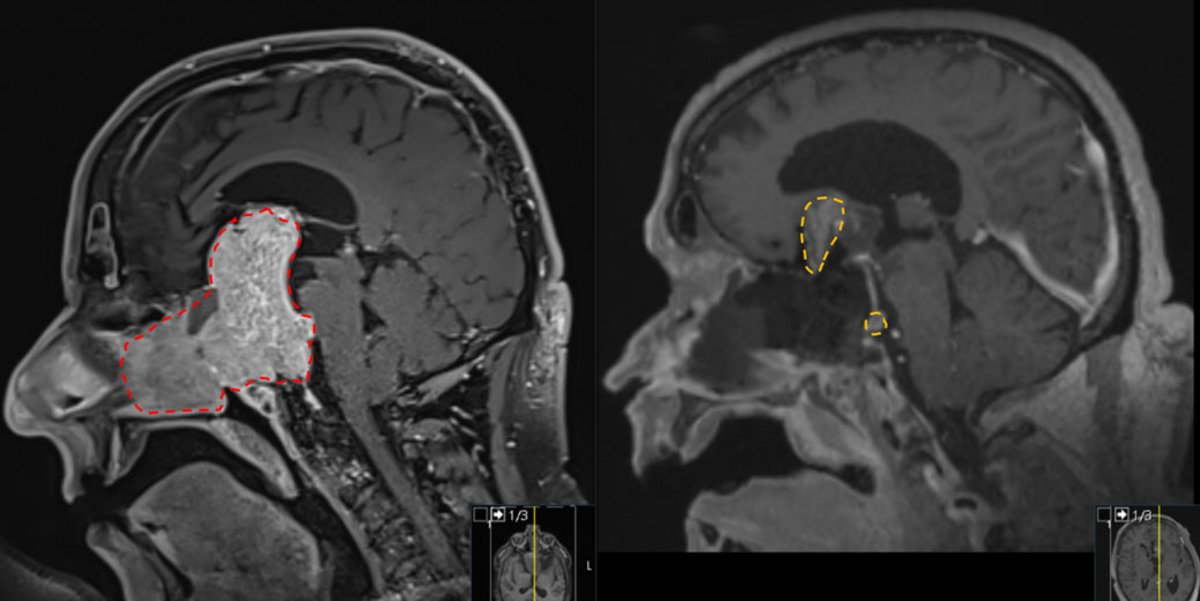
Surgical removal of the tumor is the most important form of treatment. It is particularly advisable if the tumor is damaging surrounding structures or nerves due to pressure.
Whether the tumor can be completely removed depends primarily on the experience of the surgeon, the extent to which the tumor has invaded the adjacent venous system (sinus cavernosus), and the tumor size and extent *.
For tumors with no or only limited invasion into the cavernous sinus (Knosp score ≤ 2), complete removal is successful in approximately 80% of cases. In cases of significant extension into the cavernous sinus (Knosp ≥ 3), this rate drops to approximately 30% *, *. If the tumor encases the internal carotid artery (arteria carotis interna; Knosp 4), complete removal is possible and advisable only in isolated cases.
Intraoperative MRI can play an important role in cases of large tumors or tumor components that are difficult to reach. It allows surgeons to assess, even during the operation, how completely the tumor has been removed. If necessary, the surgery can be continued immediately. This can increase the rate of complete tumor removal by 15–20% *, *.
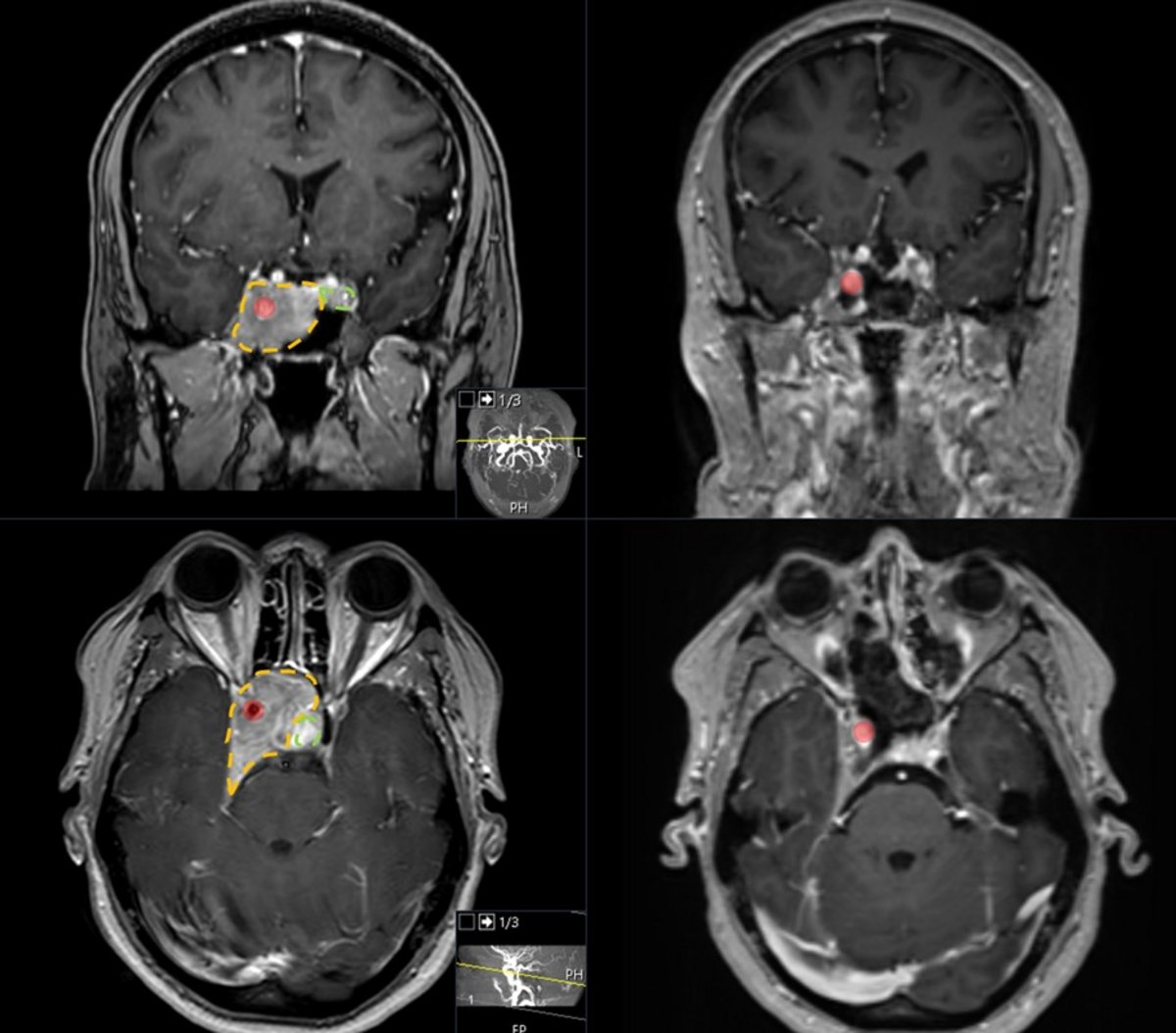
Complete tumor removal is not the goal in every surgery. If the tumor invades the cavernous sinus, a residual portion is often intentionally left in place. The reasons for this are the slightly increased risk of perforating the cavernous sinus and the high efficacy of adjunctive radiation therapy. This multimodal approach can limit the risk and enable effective tumor control.
More on the surgical technique
Radiation therapy
Radiation therapy is used as the first-line treatment only in exceptional cases. Its use is often limited by the tumor’s proximity to the optic nerve and the risk of damage to the healthy pituitary gland. However, in cases of a residual tumor or a recurrent tumor, it is an important component of a multimodal treatment plan.
Tissue examination after surgery
According to the current WHO classification, pituitary tumors are classified based on their cell lines and specific transcription factors. Pituitary adenomas are also referred to as PitNET; the abbreviation stands for «pituitary neuroendocrine tumor» *.
Approximately 75% of non-functioning pituitary adenomas are gonadotrophic at the molecular level. They express the sex hormones FSH and LH and belong to the SF-1 cell line *, *. These tumors account for about 40% of all surgically removed pituitary adenomas. Active hormone secretion is rare *, *.
In rare cases, a non-functioning pituitary adenoma corresponds at the molecular level to a silent corticotropic adenoma of the T-Pit lineage or a tumor of the Pit-1 lineage. This classification is relevant for follow-up management: A residual portion of a common gonadotrophic adenoma can generally be monitored, whereas follow-up treatment should be considered for the two rarer forms due to their somewhat more aggressive growth *.
Treatment of a hormone deficiency
A hormone deficiency must be managed by an endocrinologist. Missing hormones—particularly cortisol and thyroid hormones—are replaced with medication.
A cortisol deficiency is diagnosed based on fasting cortisol levels measured in the blood. Blood is drawn in the morning between 8 and 9 a.m. Hydrocortisone must not be taken for at least 18 hours prior to the test; otherwise, the result will be inconclusive. If the results are unclear, a Synacthen test may also be performed. A confirmed cortisol deficiency is treated with hydrocortisone. The dosage is determined on an individual basis and is usually 20–30 mg per day. To avoid metabolic and cardiovascular side effects, the lowest effective dose is selected. Affected patients should always carry an emergency ID card, as cortisol requirements increase during illness, fever, or an accident.
In cases of thyroid hormone deficiency, hormone replacement therapy is recommended; for borderline findings, close monitoring is advisable.
In cases of sex hormone deficiency, treatment with testosterone may be beneficial to improve energy levels, libido, and sexual function, as well as to maintain muscle mass and bone structure *.
ADH deficiency occurring after pituitary surgery is often temporary and requires short-term replacement therapy.
Tumor removal and further tumor monitoring
Internationally, the overall rate of complete tumor resection is approximately 50–66% *, *. For very large pituitary adenomas over 4 cm, this rate is significantly lower, at approximately 30–40 % *, *. These tumors present a complex surgical challenge and require an experienced team.
If a tumor residue remains after surgery, it continues to grow in 50–80% of cases within the following 5–10 years *, *, *, *. The larger its volume, the higher the probability of further growth *, *, *. If the residual tumor is small – less than 0.5 ml – this risk is lower *.
After complete removal, a tumor recurs within 10 years in 10–20% of cases *, *.
Regular MRI follow-ups after surgery are therefore necessary.
In the case of a growing residual tumor or a recurrent tumor, radiation therapy is usually an option. Alternatively, depending on the tumor’s location and volume as well as the patient’s condition, a second surgery may be performed. In individual cases—such as when the tumor has invaded the cavernous sinus in older patients—a planned partial resection may be a sensible approach tailored to the risk.
Hormone function
In cases of larger tumors requiring surgery, hormone deficiency is already present in approximately 56% of cases prior to surgery *.
If this deficiency is already present before the procedure, hormone function recovers after tumor removal in about 20–25% of cases *, *.
Depending on the tumor size, a hormonal axis failure occurs in 5–20% of cases following surgery *, *, *. This is often temporary but also requires medication.
Vision
If vision is impaired by the tumor, this usually leads to surgery. The prognosis for improvement is generally good. It depends on the extent and duration of the impairment, the patient’s age, and the thickness of the retinal nerve fiber layer *, *, *. The more severe and prolonged the impairment, the lower the chances of recovery.
With timely surgery, vision improves at least partially in 70–80% of cases *, *, *, *, *. Recovery can take a long time: Further improvements are possible even more than three months and up to one year after surgery *.
Why you should seek treatment at Inselspital
The treatment of non-functioning pituitary adenomas requires careful, individually tailored collaboration among various medical specialties. For complex cases, several specialized procedures are available at Inselspital:
- Endoscopic surgical technique: This technique allows access to tumor portions located far to the side or hidden, particularly when the tumor has invaded the cavernous sinus, and offers advantages over the microscopic technique.
- Intraoperative MRI: For large tumors or hard-to-reach tumor segments, the extent to which the tumor has been removed can be assessed during the procedure. If necessary, the surgery can be continued immediately. This increases the rate of complete resections by 15–20% *, *.
- Intraoperative neuromonitoring: Monitoring cranial nerve function increases the safety of the procedure.
- Multimodal treatment approach: If complete removal is not advisable, a risk-adjusted partial removal can be combined with regular follow-up visits and, if necessary, radiation therapy.
- Experienced interdisciplinary team: Very large pituitary adenomas, in particular, are surgically challenging and require an experienced team as well as concurrent care from endocrinologists and ophthalmologists.
-
Giraldi E, Allen JW, Ioachimescu AG. Pituitary Incidentalomas: Best Practices and Looking Ahead. Endocr Pract. 2023 Jan;29(1):60-68. doi: 10.1016/j.eprac.2022.10.004. Epub 2022 Oct 18.
-
Fleseriu M, Gurnell M, McCormack A et al. & The Pituitary Society International Incidentaloma Consensus Group. Pituitary incidentaloma: a Pituitary Society international consensus guideline statement. Nat Rev Endocrinol. 2025 Oct;21(10):638-655. doi: 10.1038/s41574-025-01134-8. Epub 2025 Jun 24
-
Little AS, Chicoine MR, Kelly DF, et al. & TRANSSPHER Study Group. Evaluation of Surgical Resection Goal and Its Relationship to Extent of Resection and Patient Outcomes in a Multicenter Prospective Study of Patients With Surgically Treated, Nonfunctioning Pituitary Adenomas: A Case Series. Oper Neurosurg. 2020 Jan 1;18(1):26-33. doi: 10.1093/ons/opz085. PMID: 31079156.
-
Araujo-Castro M, Acitores Cancela A, Vior C, Pascual-Corrales E, Rodríguez Berrocal V. Radiological Knosp, Revised-Knosp, and Hardy-Wilson Classifications for the Prediction of Surgical Outcomes in the Endoscopic Endonasal Surgery of Pituitary Adenomas: Study of 228 Cases. Front Oncol. 2022 Jan 20;11:807040. doi: 10.3389/fonc.2021.807040.
-
Dhandapani S, Singh H, Negm HM, Cohen S, Anand VK, Schwartz TH. Cavernous Sinus Invasion in Pituitary Adenomas: Systematic Review and Pooled Data Meta-Analysis of Radiologic Criteria and Comparison of Endoscopic and Microscopic Surgery. World Neurosurg. 2016 Dec;96:36-46. doi: 10.1016/j.wneu.2016.08.088. Epub 2016 Aug 30.
-
Hlaváč M, Knoll A, Mayer B, Braun M, Karpel-Massler G, Etzrodt-Walter G, Coburger J, Wirtz CR, Paľa A. Ten years' experience with intraoperative MRI-assisted transsphenoidal pituitary surgery. Neurosurg Focus. 2020 Jun;48(6):E14. doi: 10.3171/2020.3.FOCUS2072. PMID: 32480376.
-
Staartjes VE, Togni-Pogliorini A, Stumpo V, Serra C, Regli L. Impact of intraoperative magnetic resonance imaging on gross total resection, extent of resection, and residual tumor volume in pituitary surgery: systematic review and meta-analysis. Pituitary. 2021 Aug;24(4):644-656. doi: 10.1007/s11102-021-01147-2. Epub 2021 May 4.
-
Briggs RG, Pangal DJ, Shahrestani S, et al. Multi-Center, Multi-National Outcomes Following Endoscopic Endonasal Resection of Nonfunctional Pituitary Adenomas. J Neurol Surg B Skull Base. 2025 Mar 3;87(2):151-156. doi: 10.1055/a-2538-3928. PMID: 41808981; PMCID: PMC12971227.
-
Shinya Y, Atkinson JLD, Erickson D, Bancos I, Pinheiro Neto CD, Davidge-Pitts CJ, Peris Celda M, Herndon JS, Hong S, Van Gompel JJ. Correlation of older age with better progression-free survival despite less aggressive resection in nonfunctioning pituitary adenomas. J Neurosurg. 2024 Apr 26;141(3):781-789. doi: 10.3171/2024.1.JNS232495. PMID: 38669710.
-
Tang OY, Chen JS, Monje S, Kumarapuram S, Eloy JA, Liu JK. Comparison of Surgical Modalities for Giant Pituitary Adenoma: A Systematic Review and Meta-Analysis of 1413 Patients. Oper Neurosurg. 2025 Jan 1;28(1):1-18. doi: 10.1227/ons.0000000000001238. Epub 2024 Jul 5.
-
Cossu G, Jouanneau E, Cavallo LM, Froelich S, Starnoni D, Giammattei L, Harel E, Mazzatenta D, Bruneau M, Meling TR, Berhouma M, Chacko AG, Cornelius JF, Paraskevopoulos D, Schroeder HWS, Zazpe I, Manet R, Gardner PA, Dufour H, Cappabianca P, Daniel RT, Messerer M. Surgical management of giant pituitary neuroendocrine tumors: Meta-analysis and consensus statement on behalf of the EANS skull base section. Brain Spine. 2022 Mar 28;2:100878. doi: 10.1016/j.bas.2022.100878. PMID: 36248137; PMCID: PMC9560664.
-
Dallapiazza RF, Grober Y, Starke RM, Laws ER Jr, Jane JA Jr. Long-term results of endonasal endoscopic transsphenoidal resection of nonfunctioning pituitary macroadenomas. Neurosurgery. 2015 Jan;76(1):42-52; discussion 52-3. doi: 10.1227/NEU.0000000000000563.
-
Losa M, Mortini P, Barzaghi R, Ribotto P, Terreni MR, Marzoli SB, Pieralli S, Giovanelli M. Early results of surgery in patients with nonfunctioning pituitary adenoma and analysis of the risk of tumor recurrence. J Neurosurg. 2008 Mar;108(3):525-32. doi: 10.3171/JNS/2008/108/3/0525.
-
O'Sullivan EP, Woods C, Glynn N, Behan LA, Crowley R, O'Kelly P, Smith D, Thompson CJ, Agha A. The natural history of surgically treated but radiotherapy-naïve nonfunctioning pituitary adenomas. Clin Endocrinol (Oxf). 2009 Nov;71(5):709-14. doi: 10.1111/j.1365-2265.2009.03583.x. Epub 2009 Mar 19.
-
Hughes JD, Koeller K, Rinaldo L, Erickson D, Bancos I, Meyer FB, Atkinson J, Van Gompel JJ. Beyond Gross Total and Subtotal: Does Volumetric Resection Matter in Nonfunctioning Pituitary Macroadenomas? World Neurosurg. 2018 Aug;116:e733-e737. doi: 10.1016/j.wneu.2018.05.077. Epub 2018 May 22.
-
Panet-Raymond V, Sabit K, Shenouda G, Sirhan D, Zeitouni A, Souhami L. Growth of Pituitary Macroadenomas Postpartial Resection: Implications for Adjuvant Radiotherapy. J Neurol Surg B Skull Base. 2019 Jun;80(3):323-326. doi: 10.1055/s-0038-1670686. Epub 2018 Oct 10.
-
Seejore K, Alavi SA, Pearson SM, Robins JMW, Alromhain B, Sheikh A, Nix P, Wilson T, Orme SM, Tyagi A, Phillips N, Murray RD. Post-operative volumes following endoscopic surgery for non-functioning pituitary macroadenomas are predictive of further intervention, but not endocrine outcomes. BMC Endocr Disord. 2021 Jun 10;21(1):116. doi: 10.1186/s12902-021-00777-8.
-
Little AS, Gardner PA, Fernandez-Miranda JC, Chicoine MR, Barkhoudarian G, Prevedello DM, Yuen KCJ, Kelly DF; TRANSSPHER Study Group. Pituitary gland recovery following fully endoscopic transsphenoidal surgery for nonfunctioning pituitary adenoma: results of a prospective multicenter study. J Neurosurg. 2019 Nov 15;133(6):1732-1738. doi: 10.3171/2019.8.JNS191012.
-
Fatemi N, Dusick JR, Mattozo C, McArthur DL, Cohan P, Boscardin J, Wang C, Swerdloff RS, Kelly DF. Pituitary hormonal loss and recovery after transsphenoidal adenoma removal. Neurosurgery. 2008 Oct;63(4):709-18; discussion 718-9. doi: 10.1227/01.NEU.0000325725.77132.90.
-
Voglis S, Gönel M, Triantafyllidou M, Staartjes V, Mosca G, Bellomo J, Beyersdorf B, Akeret K, van Niftrik B, Hugelshofer M, Winzeler B, Erlic Z, Regli L, Serra C. Risk of new endocrinological deficits after intraoperative MRI-guided additional resection in endoscopic non-functioning pituitary adenoma surgery. Pituitary. 2026 Jul 11;29(4):123. doi: 10.1007/s11102-026-01727-0.
-
Fleseriu M, Hashim IA, Karavitaki N, Melmed S, Murad MH, Salvatori R, Samuels MH. Hormonal Replacement in Hypopituitarism in Adults: An Endocrine Society Clinical Practice Guideline. J Clin Endocrinol Metab. 2016 Nov;101(11):3888-3921. doi: 10.1210/jc.2016-2118. Epub 2016 Oct 13.
-
Lee DK, Sung MS, Park SW. Factors Influencing Visual Field Recovery after Transsphenoidal Resection of a Pituitary Adenoma. Korean J Ophthalmol. 2018 Dec;32(6):488-496. doi: 10.3341/kjo.2017.0094.
-
Luomaranta T, Raappana A, Saarela V, Liinamaa MJ. Factors Affecting the Visual Outcome of Pituitary Adenoma Patients Treated with Endoscopic Transsphenoidal Surgery. World Neurosurg. 2017 Sep;105:422-431. doi: 10.1016/j.wneu.2017.05.144. Epub 2017 Jun 2.
-
Müslüman AM, Cansever T, Yılmaz A, Kanat A, Oba E, Çavuşoğlu H, Sirinoğlu D, Aydın Y. Surgical results of large and giant pituitary adenomas with special consideration of ophthalmologic outcomes. World Neurosurg. 2011 Jul-Aug;76(1-2):141-8; discussion 63-6. doi: 10.1016/j.wneu.2011.02.009.
-
Pelsma ICM, Verstegen MJT, de Vries F, Notting IC, Broekman MLD, van Furth WR, Biermasz NR, Pereira AM. Quality of care evaluation in non-functioning pituitary adenoma with chiasm compression: visual outcomes and timing of intervention clinical recommendations based on a systematic literature review and cohort study. Pituitary. 2020 Aug;23(4):417-429. doi: 10.1007/s11102-020-01044-0. Erratum in: Pituitary. 2020 Aug;23(4):430-431. doi: 10.1007/s11102-020-01061-z.
-
Dekkers OM, de Keizer RJ, Roelfsema F, Vd Klaauw AA, Honkoop PJ, van Dulken H, Smit JW, Romijn JA, Pereira AM. Progressive improvement of impaired visual acuity during the first year after transsphenoidal surgery for non-functioning pituitary macroadenoma. Pituitary. 2007;10(1):61-5. doi: 10.1007/s11102-007-0007-0.
-
Asa SL, Mete O, Perry A, Osamura RY. Overview of the 2022 WHO Classification of Pituitary Tumors. Endocr Pathol. 2022 Mar;33(1):6-26. doi: 10.1007/s12022-022-09703-7. Epub 2022 Mar 15.
-
Mete O, Cintosun A, Pressman I, Asa SL. Epidemiology and biomarker profile of pituitary adenohypophysial tumors. Mod Pathol. 2018 Jun;31(6):900-909. doi: 10.1038/s41379-018-0016-8. Epub 2018 Feb 12.
-
Kim H, Kim EE, Kim YH, Park JW, Jung KC, Kim H, Won JK, Park SH. Comprehensive Classification of Surgically Resected Pituitary Neuroendocrine Tumors: Updates From a Single-Institution Experience Based on the WHO 5th Edition. J Korean Med Sci. 2025 Apr 28;40(16):e56. doi: 10.3346/jkms.2025.40.e56.